


Anaesthesia and Perioperative Care
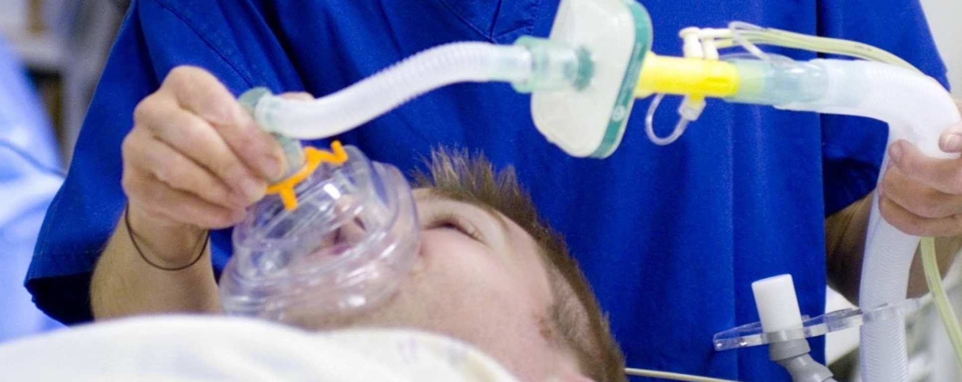 Anaesthesia makes up the largest hospital speciality and has a huge role to play in nearly every aspect of any hospital from operating theatres to accident and emergency, to the labour ward, and to intensive care. Our research ranges from basic molecular research into mechanisms of anaesthesia to investigating the clinical impact of novel anaesthetic agents.
Our research covers the entirety of patient’s perioperative journey and through this, we aim to deliver the greatest impact. The section has been pioneering in the development of novel technologies to facilitate the delivery of anaesthetic agents and has also made pivotal in-roads into the mechanism of action of anaesthetic agents and their wider application to other diseases (such as their protective roles in brain injury and in cancer).
Research themes:
- Anaesthesia mechanisms and effects
- Brain and neuroprotection
- Cardiothoracic anaesthesia and transplantation
Results
- Showing results for:
- Reset all filters
Search results
-
Journal articleBroad KD, Fierens I, Fleiss B, et al., 2015,
Inhaled 45-50% argon augments hypothermic brain protection in a piglet model of perinatal asphyxia
, Neurobiology of Disease, Vol: 87, Pages: 29-38, ISSN: 1095-953XCooling to 33.5 °C in babies with neonatal encephalopathy significantly reduces death and disability, however additional therapies are needed to maximize brain protection. Following hypoxia–ischemia we assessed whether inhaled 45–50% Argon from 2–26 h augmented hypothermia neuroprotection in a neonatal piglet model, using MRS and aEEG, which predict outcome in babies with neonatal encephalopathy, and immunohistochemistry. Following cerebral hypoxia–ischemia, 20 Newborn male Large White piglets < 40 h were randomized to: (i) Cooling (33 °C) from 2–26 h (n = 10); or (ii) Cooling and inhaled 45–50% Argon (Cooling + Argon) from 2–26 h (n = 8). Whole-brain phosphorus-31 and regional proton MRS were acquired at baseline, 24 and 48 h after hypoxia–ischemia. EEG was monitored. At 48 h after hypoxia–ischemia, cell death (TUNEL) was evaluated over 7 brain regions. There were no differences in body weight, duration of hypoxia–ischemia or insult severity; throughout the study there were no differences in heart rate, arterial blood pressure, blood biochemistry and inotrope support. Two piglets in the Cooling + Argon group were excluded. Comparing Cooling + Argon with Cooling there was preservation of whole-brain MRS ATP and PCr/Pi at 48 h after hypoxia–ischemia (p < 0.001 for both) and lower 1H MRS lactate/N acetyl aspartate in white (p = 0.03 and 0.04) but not gray matter at 24 and 48 h. EEG background recovery was faster (p < 0.01) with Cooling + Argon. An overall difference between average cell-death of Cooling versus Cooling + Argon was observed (p < 0.01); estimated cells per mm2 were 23.9 points lower (95% C.I. 7.3–40.5) for the Cooling + Argon versus Cooling. Inhaled 45–50% Argon from 2–26 h augmented hypothermic protection at 48 h after hypoxia–ischemia shown by improved brain energy metabolism on MRS, faster EEG recovery and reduced cell death on TUNEL. Argon ma
-
Conference paperWang C, Wang G, Ma D, 2015,
Pretreatment with hydrogen-enriched saline attenuates morphine tolerance
, Spring Meeting of the Anaesthetic-Research-Society (ARS), Publisher: OXFORD UNIV PRESS, Pages: E953-E953, ISSN: 0007-0912 -
Journal articleThakuria L, Davey R, Romano R, et al., 2015,
Mechanical ventilation after lung transplantation
, Journal of Critical Care, Vol: 31, Pages: 110-118, ISSN: 0883-9441Introduction: To explore the hypothesis that early ventilation strategies influence clinical outcomes in lung transplantation,we have examined our routine ventilation practices in terms of tidal volumes (Vt) and inflation pressures.Methods: A total of 124 bilateral lung transplants between 2010 and 2013 were retrospectively assigned to low(b6 mL/kg), medium (6-8 mL/kg), and high (N8 mL/kg) Vt groups based on ventilation characteristics duringthe first 6 hours after surgery. Those same 124 patients were also stratified to low-pressure (b25 cm H2O) andhigh-pressure (≥25 cm H2O) groups.Results: Eighty percent of patients were ventilated using pressure control mode. Low, medium, and high Vt wereapplied to 10%, 43%, and 47% of patients, respectively. After correcting for patients requiring extracorporeal support,there was no difference in short-term to midterm outcomes among the different Vt groups. Low inflationpressures were applied to 61% of patients, who had a shorter length of intensive care unit stay (5 vs 12 days;P = .012), higher forced expiratory volume in 1 second at 3 months (77.8% vs 60.3%; P b .001), and increased6-month survival rate (95% vs 77%; P = .008).Conclusion: Low Vt ventilation has not been fully adopted in our practice. Ventilation with higher inflation pressures,but not Vt, was significantly associated with poorer outcomes after lung transplantation.
-
Journal articleFletcher ME, Boshier PR, Wakabayashi K, et al., 2015,
Influence of glutathione-S-transferase (GST) inhibition on lung epithelial cell injury: role of oxidative stress and metabolism
, AMERICAN JOURNAL OF PHYSIOLOGY-LUNG CELLULAR AND MOLECULAR PHYSIOLOGY, Vol: 308, Pages: L1274-L1285, ISSN: 1040-0605Oxidant-mediated tissue injury is key to the pathogenesis of acute lung injury. Glutathione-S-transferases (GSTs) are important detoxifying enzymes that catalyze the conjugation of glutathione with toxic oxidant compounds and are associated with acute and chronic inflammatory lung diseases. We hypothesized that attenuation of cellular GST enzymes would augment intracellular oxidative and metabolic stress and induce lung cell injury. Treatment of murine lung epithelial cells with GST inhibitors, ethacrynic acid (EA), and caffeic acid compromised lung epithelial cell viability in a concentration-dependent manner. These inhibitors also potentiated cell injury induced by hydrogen peroxide (H2O2), tert-butyl-hydroperoxide, and hypoxia and reoxygenation (HR). SiRNA-mediated attenuation of GST-π but not GST-μ expression reduced cell viability and significantly enhanced stress (H2O2/HR)-induced injury. GST inhibitors also induced intracellular oxidative stress (measured by dihydrorhodamine 123 and dichlorofluorescein fluorescence), caused alterations in overall intracellular redox status (as evidenced by NAD+/NADH ratios), and increased protein carbonyl formation. Furthermore, the antioxidant N-acetylcysteine completely prevented EA-induced oxidative stress and cytotoxicity. Whereas EA had no effect on mitochondrial energetics, it significantly altered cellular metabolic profile. To explore the physiological impact of these cellular events, we used an ex vivo mouse-isolated perfused lung model. Supplementation of perfusate with EA markedly affected lung mechanics and significantly increased lung permeability. The results of our combined genetic, pharmacological, and metabolic studies on multiple platforms suggest the importance of GST enzymes, specifically GST-π, in the cellular and whole lung response to acute oxidative and metabolic stress. These may have important clinical implications.
-
Conference paperKoziakova M, Harris K, Campos-Pires R, et al., 2015,
The neuroprotective efficacy of noble gases in an in vitro model of ischemic brain injury.
, British Neuroscience Association, Publisher: BNA -
Conference paperCampos-Pires R, Armstrong S, Sebastiani A, et al., 2015,
Xenon provides short term & long term neuroprotection in an in vivo model of traumatic brain injury.
, BNA Festival of Neuroscience, Pages: 1-1 -
Journal articleLiu D, Zhang L, Li Z, et al., 2015,
Thinner changes of the retinal nerve fiber layer in patients with mild cognitive impairment and Alzheimer's disease
, BMC NEUROLOGY, Vol: 15- Author Web Link
- Cite
- Citations: 88
-
Journal articleBoshier PR, Mistry V, Cushnir JR, et al., 2015,
Breath metabolite response to major upper gastrointestinal surgery
, JOURNAL OF SURGICAL RESEARCH, Vol: 193, Pages: 704-712, ISSN: 0022-4804- Author Web Link
- Cite
- Citations: 4
-
Journal articleCampos-Pires R, Armstrong SP, Sebastiani A, et al., 2015,
Xenon improves neurologic outcome and reduces secondary injury following trauma in an in vivo model of traumatic brain injury
, Critical Care Medicine, Vol: 43, Pages: 149-158, ISSN: 1530-0293Objectives: To determine the neuroprotective efficacy of the inert gas xenon following traumatic brain injury and to determine whether application of xenon has a clinically relevant therapeutic time window.Design: Controlled animal study.Setting: University research laboratory.Subjects: Male C57BL/6N mice (n = 196).Interventions: Seventy-five percent xenon, 50% xenon, or 30% xenon, with 25% oxygen (balance nitrogen) treatment following mechanical brain lesion by controlled cortical impact.Measurements and Main Results: Outcome following trauma was measured using 1) functional neurologic outcome score, 2) histological measurement of contusion volume, and 3) analysis of locomotor function and gait. Our study shows that xenon treatment improves outcome following traumatic brain injury. Neurologic outcome scores were significantly (p < 0.05) better in xenon-treated groups in the early phase (24 hr) and up to 4 days after injury. Contusion volume was significantly (p < 0.05) reduced in the xenon-treated groups. Xenon treatment significantly (p < 0.05) reduced contusion volume when xenon was given 15 minutes after injury or when treatment was delayed 1 or 3 hours after injury. Neurologic outcome was significantly (p < 0.05) improved when xenon treatment was given 15 minutes or 1 hour after injury. Improvements in locomotor function (p < 0.05) were observed in the xenon-treated group, 1 month after trauma.Conclusions: These results show for the first time that xenon improves neurologic outcome and reduces contusion volume following traumatic brain injury in mice. In this model, xenon application has a therapeutic time window of up to at least 3 hours. These findings support the idea that xenon may be of benefit as a neuroprotective treatment in patients with brain trauma.
-
Journal articleCui J, Zhao H, Wang C, et al., 2015,
1 Dexmedetomidine Attenuates Oxidative Stress Induced Lung Alveolar Epithelial Cell Apoptosis <i>In Vitro</i>
, OXIDATIVE MEDICINE AND CELLULAR LONGEVITY, Vol: 2015, ISSN: 1942-0900- Author Web Link
- Cite
- Citations: 69
This data is extracted from the Web of Science and reproduced under a licence from Thomson Reuters. You may not copy or re-distribute this data in whole or in part without the written consent of the Science business of Thomson Reuters.