


Structural imaging
The MRC LMS electron microscopy (EM) facility provides equipment and expertise in the field of cryo-electron microscopy (cryo-EM), particularly single-particle structural analysis but also cellular imaging by TEM (Transmission EM) and SEM (Scanning EM).
All the stages in project development preceding high-resolution data collection, on a high-end microscope, can be performed at the EM facility. Equipped with a new Talos F200i TEM, a Falcon 3EC direct electron detector, a CETA camera as well as ELSA and 910.6 cryo-holders, the users can analyse negatively-stained samples and collect preliminary cryo-EM data to get sub-nanometric structures. Users have also access to a Vitrobot Mark IV and a Leica EM GP2 plungers for cryo-EM sample preparation, a Cressington 208 high-vacuum carbon coater and a Pelco easiGlow unit to glow discharge grids.
We also have equipment for ultrathin sectioning of resin-embedded cellular specimens. They can be prepared and imaged at the facility using the Leica EM ACE 600 sputter coater, the Ultramicrotome UC7 with the Talos F200i for high-resolution transmission imaging, or with the Apreo Volumescope SEM for serial block-face imaging. Users can also be guided into accessing technologies in neighbouring facilities as part of the development of the new correlated light and electron microscopy (CLEM) capabilities at the EM facility and the Leica Thunder Imager cryo-CLEM.
The facility provides also training, advice and help for data collection and image processing for structure determination. The facility has a dedicated GPU workstation for data processing.
Equipment images
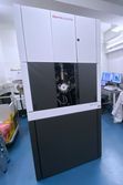
Thermo Fisher Talos F200i TEM
Thermo Fisher Talos F200i TEM

Thermo Fisher Apreo Volumescope SEM
Thermo Fisher Apreo Volumescope SEM
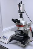
Leica UC7 ultramicrotome
An image of a Leica UC7 ultramicrotome used in structural Imaging

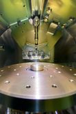
Block trimming with a diamond knife
Block trimming with a diamond knife
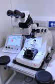
Leica Thunder Imager cryo-CLEM
Leica Thunder Imager cryo-CLEM
Leica ACE600 gold sputtering
Leica ACE600 gold sputtering