Imperial academic talks about studying museum specimens of Chinese bound feet
by Colin Smith
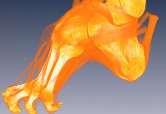
A 3D model of a bound foot
An Imperial academic talks about her recent work in analysing the internal structure of bound-feet museum artefacts.

A bound foot (Credit: Jo Farrell Photography)
Dr Natalie Reznikov, from the Department of Materials at Imperial College London, has been analysing the specimens of bound feet, which are more than 170 years old. Her research is published today in the Journal of Bone and Mineral Research .
Dr Reznikov talks to Colin Smith about the insights these cultural relics are providing her about bone structure.
What is foot binding?
For nearly a thousand years in China, women underwent foot binding to achieve the “prize” of a smaller foot, which was considered the height of fashionable beauty and aristocratic refinement. It was a cultural practice where the foot was reshaped by restraining growth and creating what was called the tiny ‘golden lotus’ shape. The consequences of this practice were that millions of women were crippled.
Can you explain the binding process?
The process started at a very early age when a girl was six or seven. Foot binding involved using ribbons of silk or cotton to bend most of the toes under the foot, except the big toe. More fabric was used to compress the heel bone and the ball of the foot together. Over several years, the foot was squashed into a smaller shape.
What were the physiological consequences of this process?

Dr Reznikov
The realignment of the foot bones meant that the foot no longer provided a shock dampening function, which we take for granted. These women often struggled to walk, taking much smaller swaying strides, which was thought attractive. The stiff and restrained feet meant that the women had to balance their weight mainly on their heels. They may have even developed much thicker fat pads on the heels to compensate for the changes in pressure on their bones. There was also a range of side effects in their feet such as pain, poor blood circulation and ulcers. Foot binding was eventually outlawed in China in 1912 by the founder of the Chinese republic, Sun Yat Sen, following the overthrow of the last Qing Dynasty emperor of China.
Where did the bound feet artefacts you analysed come from?
They were originally brought back to the UK by a naval surgeon called Steven Stanley, sometime after 1842, following the first Anglo/Chinese War. Stanley died on an expedition following his return from China and the artefacts were donated by his wife to the Royal College of Surgeons of England. They kindly lent them to me to carry out my research.
Why did you study them?

The architecture of the spongy material inside the bone was modeled by Dr Reznikov
Analysing the shapes of bones tells us a lot about their function and how they were used by their owners. For example, bigger bones in one arm may indicate to an osteoarchaeologist that the person may have favoured one arm more than the other. This could suggest the individual was involved in repetitive work, perhaps using a tool such as a hammer constantly.
However, it has always been difficult to separate which features in bones are a product of evolution and which features in the bone are adaptations that have happened over a person’s lifetime.
The bones in the bound feet are really useful to explore this issue. That is because they’ve been completely adapted for another purpose. By comparing them to healthy feet I hoped to see which features in bone happened through evolution and which features were the caused by adaptation.
What did you look at?
The interior of the heel is made up of a spongy, delicate network of struts called the trabeculae. We analysed it. We compared what we found in the bound feet to the healthy feet.
The specimens were scanned at the Natural History Museum using a computed tomography (CT) scanner, which uses X-rays to make detailed pictures of the feet. In all, we took several thousand projections of each specimen, which we then collated into 3D digital models of the heel bones.
What did you discover?
Firstly, we discovered an identical blueprint associated with stability and strength in the heel in the in both the bound and healthy feet. This implies this blueprint of spongy bone is an evolutionary trait.
Secondly, we found that the part of the spongy network of struts that have to cope with stresses and strains from the lower leg and tendons were different in the healthy and bound feet.
The 3D patterns in the healthy heel reflected the role it played as a shock absorber. In the bound heel, the pattern of struts was a rigid pillar, reflecting how the foot was more of an extension of the lower leg in people with bound feet.
This suggests that the local organisation and shape of the network of struts is an adaptive trait that evolves during a person’s life.
Do you think your 3D modelling approach could be used in any other fields of research?
We believe the spongy bone struts are like microscopic signposts that encode the biomechanical history of an individual. It could be of benefit for archaeologists, anthropologists and evolutionary biologists looking to reconstruct past lifestyles or evolutionary processes by investigating the inner architecture of bones. The research also opens new insights into medical research, for example, sports medicine or orthopaedics.
Article text (excluding photos or graphics) © Imperial College London.
Photos and graphics subject to third party copyright used with permission or © Imperial College London.
Reporter
Colin Smith
Communications and Public Affairs