Location, location, location: how location effects the structure of heart cells
The muscle cells in your heart respond differently to adrenaline depending on where they are found due to underlying structural differences.
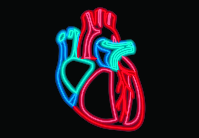
You may be forgiven for thinking that one heart cell is pretty much the same as the next. However, a recent study from Professor Julia Gorelik and Dr Peter Wright has discovered that not every heart cell is created equal. Their research looks at cardiomyocytes, the muscle cells of the heart, which are responsible for pumping the blood from our heart through our bodies. They’ve uncovered structural differences in these cells dependant on where they are located in the heart, which could have far reaching implications for other research into heart disease.
“The cardiomyocyte is possibly the most interesting cell in nature because of what it has to do” Dr Peter Wright First author
Peter and Julia initially decided to investigate these cells to better understand the disease mechanisms behind Takotsubo cardiomyopathy, commonly known as ‘broken heart syndrome’. Takotsubo is characterized by a change in the contraction of the heart, whereby the apex of the heart (at the bottom) appears to lose contractility relative to the ‘base’ (found at the top of the heart). It has been suggested that this is because the cardiomyocytes in the apex respond to adrenaline differently to those in the base. Julia and Peter wanted to understand whether the receptors which help the cardiomyocytes interpret adrenaline and other similar signalling molecules, were acting differently in different parts of the heart. People consider all heart cells to be the same but clearly there was something different about the cells of the left ventricle in the apex of the heart, which gave rise to the disease. They decided to take a closer look, comparing cells from different parts of the heart. What they have found is that the root of these differences is that the structure of the cells is different. They also concluded that cardiomyocytes in different areas of the heart react differently to adrenaline.
The researchers began by looking at differences in regional properties in cells from around the heart and they found that the response to drugs, and the activity of cells, was different. Further investigation found that the morphology of the cells in different parts of the heart is quite different which effects how they respond to stimulation by adrenaline. The group are particularly interested in receptor signalling and this study looks at Beta-2-adrenergic receptor, one of three beta adrenaline receptors found in the heart. The study found that differences in structure mean that the same adrenaline signal will be interpreted differently by cells located in different parts of the heart. The same amount of adrenaline will cause more contraction in the apex of the heart (at the bottom) than the basal ones (at the top). Basal cells have a higher degree of structural organization and as a result the adrenergic receptors at the top of the heart are more controlled, or ‘compartmentalized’, compared to the apex of the heart. This affects the control of signalling by the receptors. Less structural organization equals a larger amount of signalling by the receptor in the apex of the heart. Adrenaline signalling is primarily important in the heart’s ‘fight or flight’ response as we respond to an imminent threat.
This work demonstrates another factor which may contribute to why Takotsubo occurs. A small amount of adrenaline enhances heart function acutely, but a lot of adrenaline can damage cells. Takotsubo is another condition where we see our biology stretched to its limits, when usefully evolved structures are pushed beyond their normal function, they can start to go wrong.
Gorelik and Wright’s work also has wider implications for others studying the heart. Cardiology researchers often isolate heart cells to study them to better understand how the heart functions, what causes disease and how this can be treated. The knowledge that the same cell type, taken from a different part of the same heart, may give different results could be vitally important for ensuring other research can provide us with accurate answers. Usually researchers take a group of cells without being selective as to where in the heart they come from, run their tests and then look for statistical significance in their results. This study found that there are subtle structural differences between areas of the heart, which probably gives rise to the efficient working of heart as it is. Other researchers doing work in this area maybe missing an important piece of the puzzle with regards how the heart’s structure is built up by ignoring these differences between cells.
Many groups are creating computational models of how the heart functions, recreating what they think happens in a heart cell and multiplying it to build a model to simulate what happens in the heart as a whole. But any model is only as good as the assumptions it is built on, and if the assumption is the cells are all like simple Lego bricks and you just connect them together, this is wrong. They cells are not like plain Lego bricks, but instead more like smart bricks that are all different and interacting with each other. Furthermore researchers are developing synthetic constructs to develop damaged parts of hearts with tissue engineered from stem cells. The findings of this study may be useful in that they may stimulate other work to ensure that engineers are replacing like for like, rather than generic tissue. The heart’s structure develops as we grow, and the genetic information and environmental pressures lead to the formation of a unique cell. We need to carry on dissecting and decoding how these structures form and then work, to better inform future translational work.
Why are there these differences in the structure of cells in the healthy heart? The working hypothesis is that due to shape of the heart there are different stress demands on different areas. Its cone shape allows it to create and push a bolus of blood through your body as it pumps and twists. But this difference in radius through the chamber will translate into different strain on the muscle cells located in different areas. So, the researchers postulate that the reason for the different shape and responsiveness of cells is because they are put under different demands. The next step is to confirm what factor is controlling the difference in cells between apex and base. The team have been using a novel model to allow them to manipulate the load on cardiac tissue to try and find out what fundamentally gives rise to the heart’s structure.
Every cell that makes up our bodies is part of an environment that is dynamic and varying. This study raises the potential importance of variation of cells in different parts of the same organ, and subsequent effects of signalling processes. The idea that cells of a similar type can exhibit marked structural variation is not a particularly new one (Ramon y Cajal, proposed that all neurones were unique, in the 19th century). This study shows this principle can be applied more widely to the body’s organs. All our organs, and their cells, are under various strains and forces that affect them over time. Previous work by Julia’s group has shown that the endothelial cells that line our blood vessels change their shape depending on what part of the vessel they are on. This is due to the pressure, stress and shear force of the blood, which they must endure. In the aortic arch the cells along the top are long thin and grooved, whereas the bottom cells, where there is a slower flow of blood are more like cobble stones.
Genetics leads to cell division and organisation of cells into organ structures but environmental pressure and forces then shape them further. This study raises important questions regarding the degree to which a cell’s shape and properties are determined by environmental rather than genetic factors.
Article supporters
Article text (excluding photos or graphics) © Imperial College London.
Photos and graphics subject to third party copyright used with permission or © Imperial College London.
Reporter
Ms Helen Johnson
Communications Division