Chemical imaging used to diagnose colon cancer
by Sara West
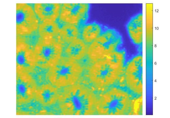
Fourier-transform infrared (FTIR) spectroscopic imaging of colon tissue
Researchers have developed a method of chemical imaging which identifies colon cancer more accurately and efficiently than traditional methods.
Colon cancer is the fourth most common cancer in the UK, causing 16,000 deaths each year. Early intervention ensures the highest chance of survival, but symptoms can often be mistaken for other illnesses.
This new method uses computer analysis to improve infrared imaging techniques and produce more accurate results, paving the way for clinicians to diagnose patients more efficiently.
“Chemical photographs”
The research team in the Department of Chemical Engineering at Imperial College London used Fourier-transform infrared (FTIR) spectroscopic imaging to produce ‘chemical photographs’ of biopsy tissue samples ranging from heathy to cancerous.
FTIR imaging involves shining an infrared beam at a sample and measuring how much of that light is absorbed at different frequencies, which is used to produce a visual reference of the sample’s chemical composition.
The results encourage us to transfer this technique to actual clinical settings to improve disease detection. Ms Cai Li Song Lead Researcher
The results, published in Analytical and Bioanalytical Chemistry (Springer Nature), show significant chemical differences in the samples at different stages of disease. This is important because cell changes take place on a chemical level before any physical deformities occur, enabling clinicians to detect changes early on.
This study demonstrates the value of using FTIR spectroscopic imaging as a diagnostic tool for colon cancer, alongside tools such as colonoscopy, surgery and histopathology.
Producing predictive models
The researchers also used a random forest (RF) classifier programme to analyse the data from the spectroscopic image. In doing so, they demonstrated for the first time that only the fingerprint region of the mid infrared spectrum (7-10 micrometers) is important when diagnosing cancerous malignancy.
This is significant because data taken from a wider range of the spectrum is at greater of risk of distortion from Mie scattering, where light particles scatter, which can only be fixed with a corrective lens or a complex computer algorithm.
By only using the data from the fingerprint region each of these lengthy processes can be eliminated, and equipment training for clinicians made less time-intensive.
Application in clinical settings
Although this study was restricted to detecting colon cancer, the researchers have used the results to create models which have the potential to be applied other difficult to diagnose cancers such as oesophageal cancer, and even-non cancerous anomalies.
Lead researcher Ms. Cai Li Song said: “We demonstrated a label-free digital pathology by infrared spectroscopic imaging technique with subsequent chemometrics to allow us to differentiate benign and malignant colon polyps. The results encourage us to transfer this technique to actual clinical settings to improve disease detection.”
Professor Sergei Kazarian added: “There is urgency in developing new techniques, which can identify the early stages of cancer in a way that goes beyond the current histopathology approaches in order to increase survival rates.
Coupling spectroscopic imaging with advanced machine learning approaches aid will early detection and understanding of cancer. There is an excitement of having an enhanced accuracy that promises advances in the early cancer detection and differentiation of disease stages.
Cai Li Song, an outstanding PhD researcher in my group, has obtained the spectral data of high-quality and applied innovative selection of spectral features to colon cancer classification with high accuracy, thus bringing spectroscopic imaging closer towards clinical acceptance.”
Notes
The full paper is available online via Open Access.
Tissue samples were provided by Prof. R. Goldin at St Mary’s Hospital, Imperial College London.
Article text (excluding photos or graphics) © Imperial College London.
Photos and graphics subject to third party copyright used with permission or © Imperial College London.
Reporter
Sara West
Communications Division