This project is focused on developing structural finite element models of the lumbar vertebrae. Based on the iterative adaptation method developed in the group, skeletal models of the vertebrae are optimised to different mechanical loading envelopes.
The external geometries of the lumbar vertebrae are segmented from the same high-resolution MRI scans of healthy volunteers as the musculoskeletal model of the lumbar spine. Using the method applied for the mesoscale structural model of the femur, a volume mesh consisting of tetrahedral elements is obtained. The surface nodes of the mesh are used to create shell elements representing cortical bone, whilst truss elements are generated by pairing nodes within the volume to represent trabecular bone.
Experimentally obtained loading conditions using the London Lumbar Spine Model are applied to the finite element models. Thicknesses of cortical shells and radii of trabecular trusses are iteratively adapted to achieve a target strain when resisting the loading envelope.
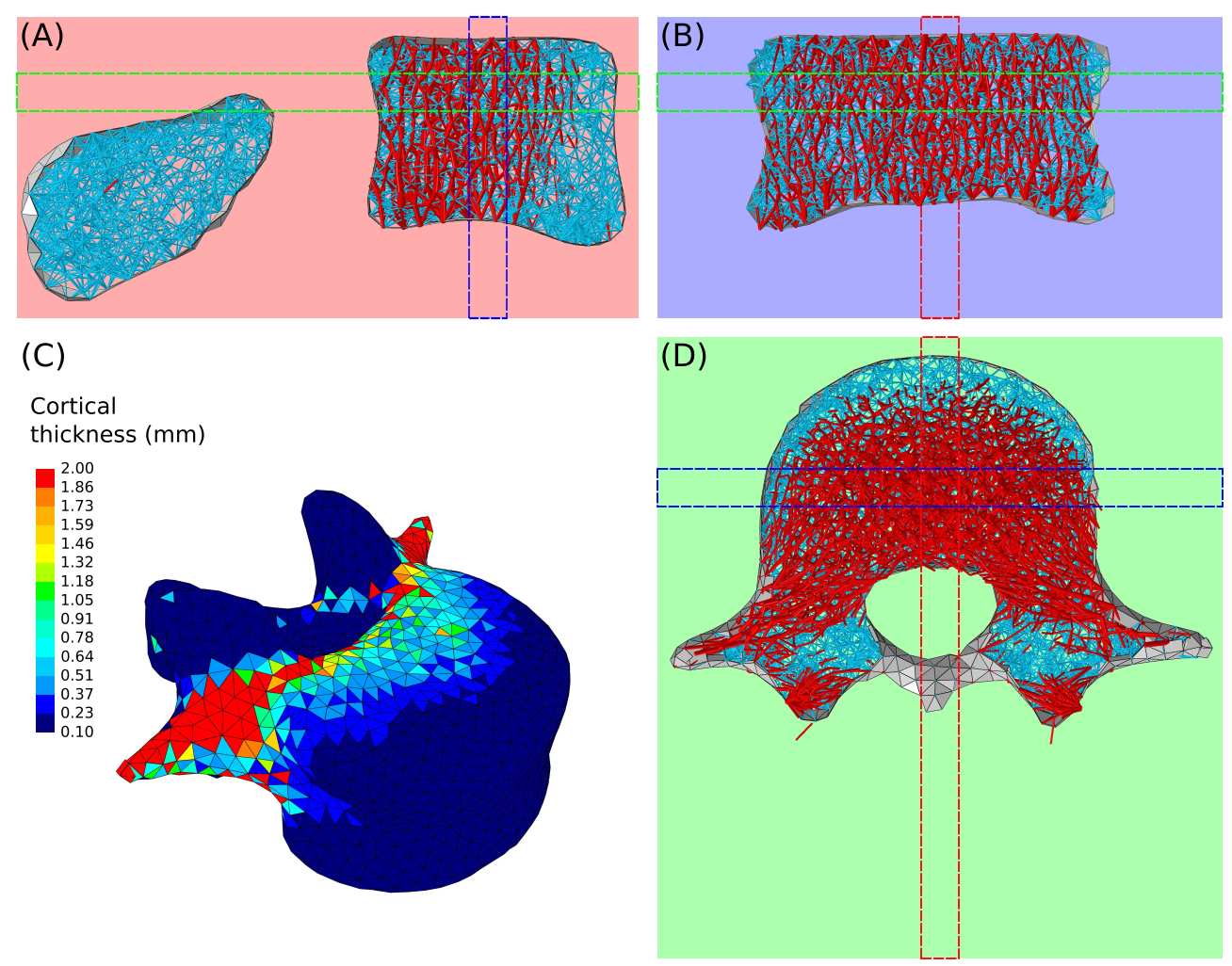
Structural architecture of L4 adapted to a healthy loading envelope. (A) midsagital, (B) midcoronal and (D) through processes transverse slices showing the internal architecture. Cortical bone is displayed in grey, trabecular bone in red. Trabecular truss elements with radius <= 0.1mm are shown in blue. (C) Cortical thickness ranging from 0.1 to 2mm.
Healthy loading scenario
A loading envelope representative of a healthy lifestyle is obtained from muskuloskeletal simulations for a range of activities of daily living. These activities include locomotion activities involving the lower limbs (walking, stair ascent and descent, sit-to-stand and stand-to-sit), small spine movements limited to 20° (flexion, extension , lateral bending and axial rotation), and large spine movements (maximum flexion to reach toes, lacing shoes while sitting, range of lifting tasks involving twisting of the spine from sitting or standing positions). Adaptation of the structural finite element models of the lumbar vertebrae under this loading envelope predicts the bone architecture characteristic of the vertebral inner structure: trabecular trusses resisting compression forces in the vertebral body and tension in the transverse processes, thicker cortex resisting bending in the pedicles and transverse processes.
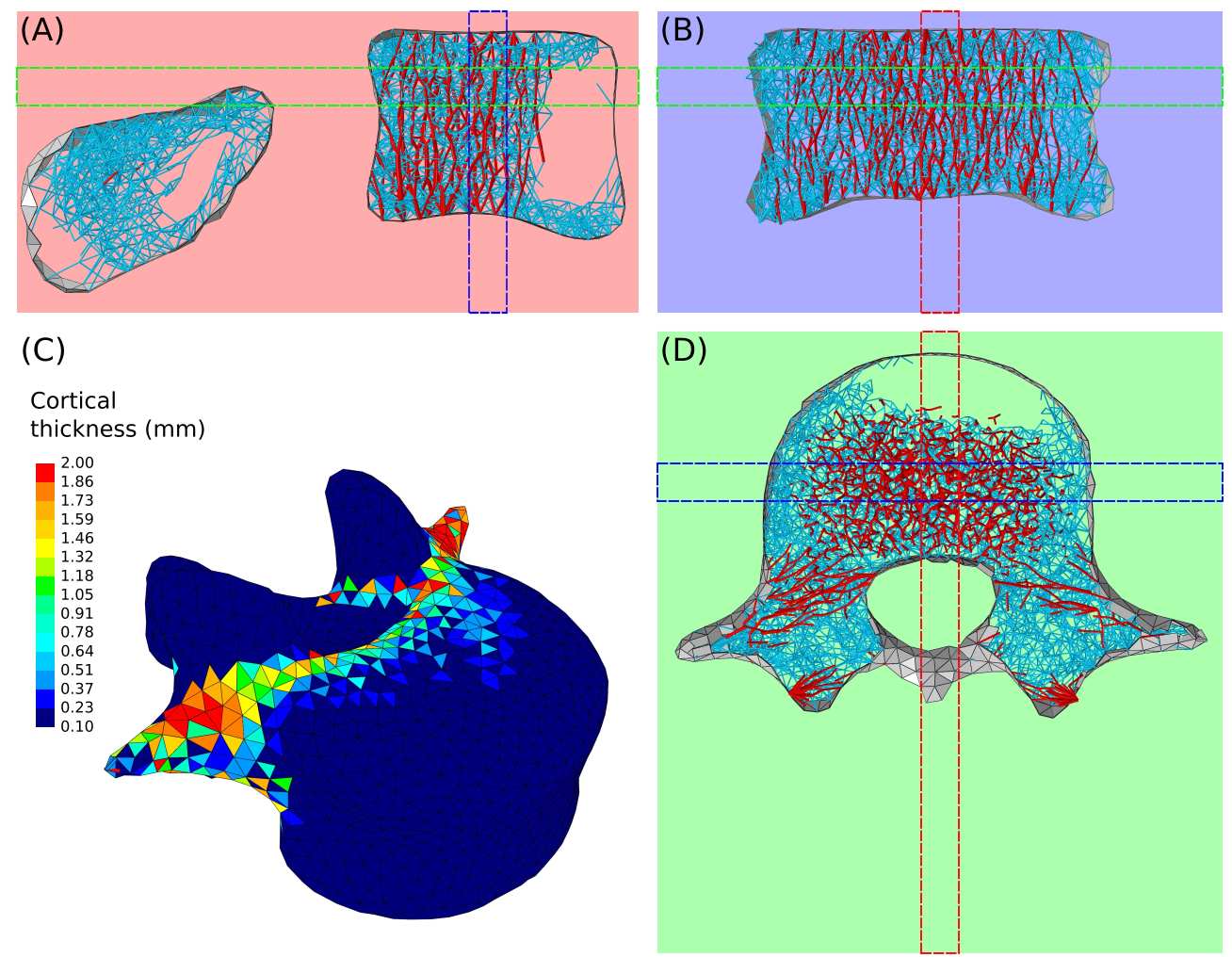
Structural architecture of L4 adapted to an altered loading envelope without lifting tasks and large spine movements. (A) midsagital, (B) midcoronal and (D) through processes transverse slices showing the internal architecture. Cortical bone is displayed in grey, trabecular bone in red. Trabecular truss elements with radius <= 0.1mm are shown in blue. (C) Cortical thickness ranging from 0.1 to 2mm.
Altered loading scenario
The modelling framework can also be used to predict bone adaptation to altered loading conditions. The altered loading scenario presented here is composed of the locomotion activities and small spine movements described in the healthy scenario. Lifting tasks and large spine movements are not included. With these altered loading conditions, the adapted structures of the lumbar vertebrae lack several features that are normally observed in healthy vertebrae. Cortical bone is thinner in the pedicles and transverse processes. Trabecular bone is also less dense and has even disappeared in the anterior part of the vertebral body and the spinous process.
The modelling framework is a powerful tool which can be used to predict bone architecure in different loading scenarios representative of healthy and pathological conditions. Future work will focus on applying this computational approach to clinical scenarios and help refining specific treatments and guidelines to maintain lumbar spine bone health.