Feed or flee
The brain cells that tell us when to eat and when to run away

Over time, too little food can leave us with stunted growth and an inability to fight off infections. Too much food can lead to obesity, raising the risk for diabetes, cardiovascular disease and cancer.
But while the need to feed is one of our most basic biological processes, the networks of brain cells which control it are turning out to be far more complex than we imagined.
Increasingly, the evidence suggests that the nerve signals fired from the same cluster of neurons that tell us when it’s okay to eat, also help us flee from danger and run away to feed another day.
Scientists believe that a better understanding of how this core process is controlled could help in the development of new treatments for obesity and psychiatric conditions linked to anxiety.
While the traditional view may have seen the regulation of emotions and appetite as two separate streams of the same system, the evidence has been mounting – you can’t target one without having an impact on the other.
Switching behaviours

“For animals in the wild we know that feeding is a risky and often dangerous undertaking, and that’s always been the case,” explains Professor Dominic Withers, from Imperial’s Institute of Clinical Sciences / MRC London Institute of Medical Sciences.
As a clinician, Withers’ interests lie in obesity and diabetes. But understanding how the urge to eat is controlled has taken him back to the lab to look at the neurological basis of feeding in mice.
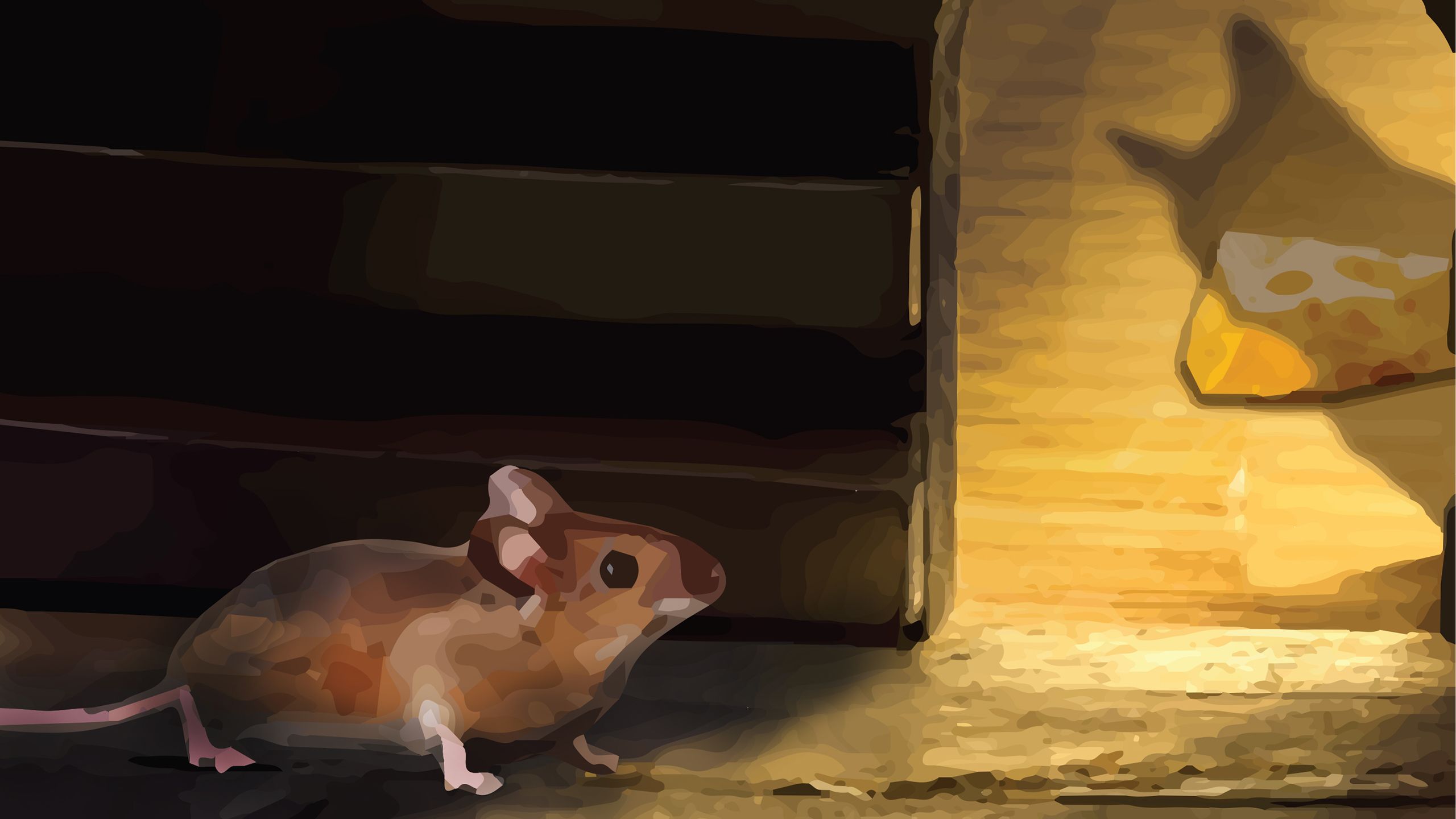
You go out to feed and you could just as easily become food yourself...so the ability to suddenly switch to defensive behaviour is important"
Professor Withers explains that animals need to integrate their exploration for food, and other behaviours associated with feeding, with the ability to run away and escape.
When investigating obesity one region of the brain in particular – the ventromedial hypothalamus (VMH) – has been a focus of research for decades. This cluster of neurons, which lies deep in the brain within the hypothalamus, is known to drive behaviours around feeding, mating and aggression.
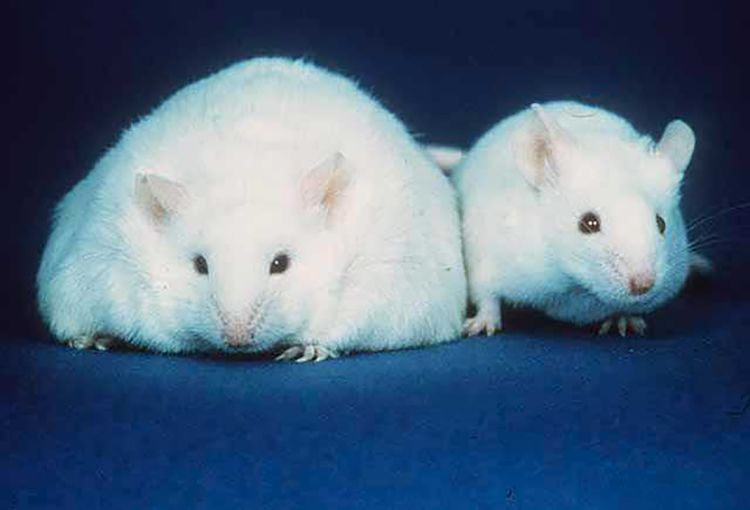
Previous studies have shown that when the VMH is damaged in rats, they direct the majority of their energy towards eating or fighting, leading to animals with massive weight gain and increased aggression between males fighting over females.
Exactly how this cluster of cells switches between the drive to fight, run away, mate or feed, has remained unclear. But work from Withers' team has revealed a complex interplay between distinct groups of cells.
Fine Tuning Feedback


In order to understand more about eating disorders in humans the Imperial team has focused on rodents, which have the same regulatory networks in their brains.
“People have studied animals with damage to the VMH and seen profound effects on bodyweight and feeding, but they didn't actually know which circuits they were altering when they damaged this region,” he explains. “But our work provides very discreet details and defines precisely what population of neurons is making you feed and making you not eat.”
Drawing on cutting-edge neuroscience tools, the team has been able to peer inside the brain to reveal what happens to these neurons during feeding, revealing another layer of control in a subset of cells in the VMH.
The researchers turned to optogenetics, using mice with genetically modified neurons that can be stimulated with flashes of laser light, enabling them to control the activity of specific regions of the brain with pinpoint accuracy.
Honing in on the VMH, they found a distinct population of cells called SF1 neurons, which act as a fine control.
Under normal conditions, these cells fizz with activity as the animals explore their surroundings, maintaining a baseline level of anxiety in the animals. But when the animals approach food their behaviour needs to change, to drop their guard slightly while they eat.
Using specialist miniature cameras mounted on the animals’ heads and monitoring the flow of calcium ions in and out of cells, the team was able to record the activity of neurons in real time, revealing changes in the activity of SF1 when the animals approach food.

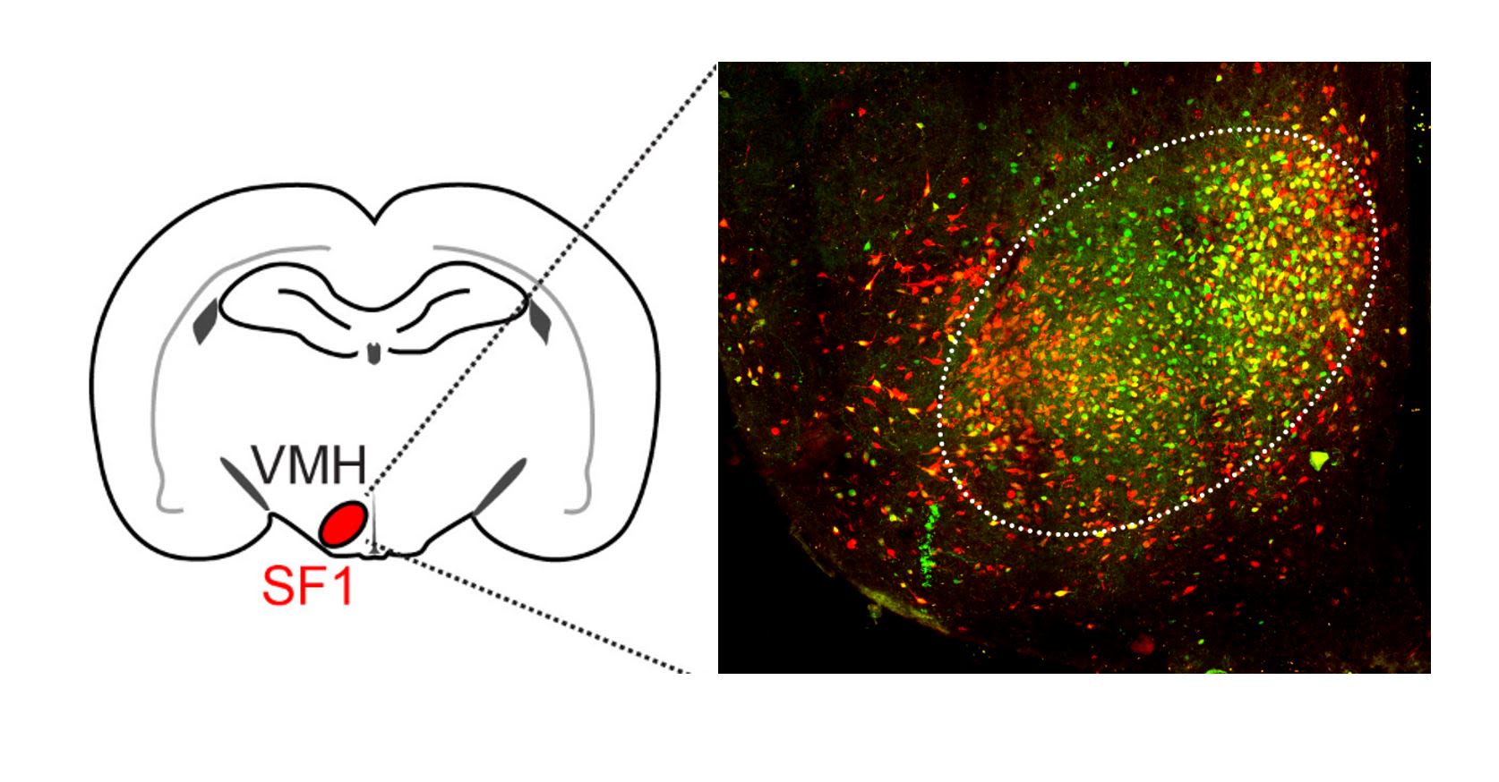
A population of cells within the VMH (called SF1) act to fine tune the urge to feed
A population of cells within the VMH (called SF1) act to fine tune the urge to feed
Just as the mice drop their guard temporarily to feed, the activity of the cells dampened down while the animals ate, enabling the VMH to switch to the 'need to feed'. But activity increased again after feeding had stopped. The findings indicate that the SF1 are acting as the gatekeepers for this switch, carefully regulating the animals’ behaviour.
“The minute you up the activity in this neural region, the mouse switches from eating to defensive behaviour to make sure it's safe,” explains Paulius Viskaitis, a PhD student in the Withers Lab who undertook much of the work.
“It is quite clear that SF1 aren't self-contained, they talk to many brain regions, so act as a gatekeeper to activate those other systems," adds Viskaitis. "These circuits are becoming bewilderingly complex, in terms of how they are all wired together and what is permitted and what's not.”
Less food, more risk?


The team found that feeding behaviour could be influenced by manipulating the activity of the SF1 and upping the anxiety levels of the mice. By making the mice more anxious (through increasing their stress levels) they were able to effectively ‘flick a switch’ inside their brains, shutting down the desire to feed.
Animals given drugs to make their SF1 neurons fire more often (so were more anxious as a result) were less likely to feed and stored less fat. In contrast, suppressing the activity of the SF1 decreased anxiety and made mice eat more and gain weight.
Intriguingly, the evidence suggests that these gatekeeper cells are also sensitive to the nutritional status of mice – how well fed they are. In animals that received just 80% of their normal food intake, the pressure to eat would override the anxiety. When the SF1 neurons were stimulated with light, increasing their activity, the avoidance effect was blunted in these mice, leading to them taking more risks to eat.
We have shown for first time that activity in this small population of brain cells acutely changes food intake"
The work is painting a picture of complex networks of brain clusters, all of which feed-back to one another to carefully regulate feeding. But what can this tell us about our own health?
The human link


According to the team, beyond the lab, the findings could have implications for human eating disorders and stress.
“There’s a long-standing recognition that things like obesity are associated with altered anxiety states and altered emotions and depression, so it is a bit of a chicken and egg as to which came first,” said Withers.
He describes a “litany of failures” in the path to develop psychiatric treatments and medications for obesity, with many abandoned due to side effects.
In 2008, rimonabant, a drug licensed for the treatment of obesity, was pulled from the market following reports of serious psychiatric effects among patients.
Similarly, a number of antipsychotic treatments have been linked with weight gain in patients, typically in the first six months of treatment, and weight related side effects have been reported with common antidepressants and anti-anxiety medications.
“The circuits and chemical transmitters involved in feeding and emotions overlap quite significantly,” explains Withers, “so that's one of the reasons why it's so difficult to target those conditions without hitting pathways that affect the other – it’s difficult to disentangle psychiatric and metabolic drugs and vice versa.”
He adds that small molecule drugs may hold the potential to unlocking this, by targeting the brain’s fine control mechanisms, such as the SF1 neurons, rather than the approach of current drugs which cast the net far wider to have an effect, but increase the risk of unintended side effects.
The turning point may be the availability of cutting edge tools for accurately monitoring and altering the activity of specific brain regions.
When you start combining these new tools in the lab, we're really moving into a revolution in brain science"
Moving beyond the traditional simplified model of the brain may be the key to overcoming some of the previous failures, offering hope for patients with metabolic and psychiatric conditions alike.
“At the moment we're only in the foothills of discovering how the brain works, particularly the appetite regulatory circuits,” he adds. “But when you start combining these new tools in the lab, we're really moving into a revolution in brain science.”


An MRI scan of the human brain
An MRI scan of the human brain
