Microscopy and Imaging
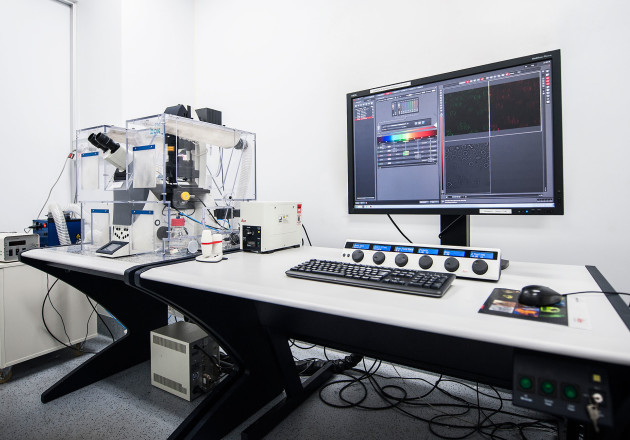
The microscopy and imaging facility provides users with access to high specification widefield, confocal, light and fluorescence microscopes, as well as a state-of-the-art micro-CT scanner. The facility is spread across both the South Kensington and White City campuses and includes the specialist Imperial College London and Leica Microsystems Imaging Hub.
The imaging equipment is available to all members of the department plus those from the wider Imperial College community and beyond.
You will also be provided with the expertise of dedicated imaging specialists who will be happy to assist you with equipment training, experimental design, image acquisition and analysis.
All users must be trained by the facilities’ dedicated technical staff before using the imaging equipment. Bookings and training requests are made via PPMS.
Locations: Bessemer (South Kensington) and Sir Michael Uren Hub (White City).
Further information can be found on the imaging facilities' dedicated Wiki pages.
Available Imaging Equipment - South Kensington
Leica SP8 Lightning Confocal Microscope
Specifications
- Microscope
- DMi8CEL inverted
- Scanning stage
- Universal insert
- Microplate insert
- LED illumination with DAPI, FITC and RHOD filters
- Lasers
- 405 UV laser
- Super continuum white light laser (470-670nm)
- Objectives
|
Correction
|
Magnification
|
NA
|
Immersion
|
Coverglass
|
Working distance
|
Other
|
|---|---|---|---|---|---|---|
| HC Plan APO | 10x | 0.40 | Dry | 0.17 | 2740um |
CS2 |
| HC Fluotar L VISIR | 16x | 0.60 | IMM incl BABB | - | 2500um | Correction ring (suitable for optically cleared samples using BABB (aka Murray's clear) |
| HC Plan APO | 20x | 0.75 | Dry | 0.17 | 620um | CS2 |
| HC Plan APO | 40x | 1.30 | Oil | 0.17 | 240um | CS2 |
|
HC Plan APO |
63x | 1.40 | Oil | 0.17 | 140um | CS2 |
- Detectors
- 1x PMT
- 2x HyD
- Environmental control
- Temperature
- Extras
- Lightning
Leica SP5 Confocal Microscope
Specifications
- Microscope
- DMi 6000CS inverted
- Scanning stage
- Universal insert
- Microplate insert
- SuperZ galvo
- Mercury lamp illumination with DAPI, FITC and RHOD filters
- Lasers
- Argon: 458nm, 476nm, 488 nm, 496nm and 514nm
- DPSS: 561nm
- HeNe: 633nm
- Objectives
- PL APO PH 10x/0.40 dry (WD 2.2mm)
- PL APO 20x/0.70 IMM (WD 0.26mm water - 0.17mm oil)
- PL APO PH 40x/1.25 oil (0.1mm)
- PL APO 63x/1.3 Glycerol (0.28mm)
|
Correction
|
Magnification
|
NA
|
Immersion
|
Coverglass
|
Working distance
|
Other
|
|---|---|---|---|---|---|---|
| HC Plan APO | 10x | 0.40 | Dry | 0.17 | 2200um |
CS |
| HC Plan APO | 20x | 0.70 | IMM | 260um (water) - 170um (oil) | Lbd. Bl | |
| HCX Plan APO | 40x | 1.25 | Oil | 0.17 | 100um | CS |
| HCX Plan APO | 63x | 1.40 | Oil | 0.17 | 100um | Lbd. Bl |
|
HCX Plan APO |
63x | 1.30 | Glycerol | 0.14-0.18 | 280um | CS |
- Detectors
- 3x PMT
- Environmental control
- Temperature
Light and fluorescence microscopes
|
Light microscopes
|
Make/Model |
Features |
 |
Leica DM IL |
|
|
|
Leica DM IL |
|
|
|
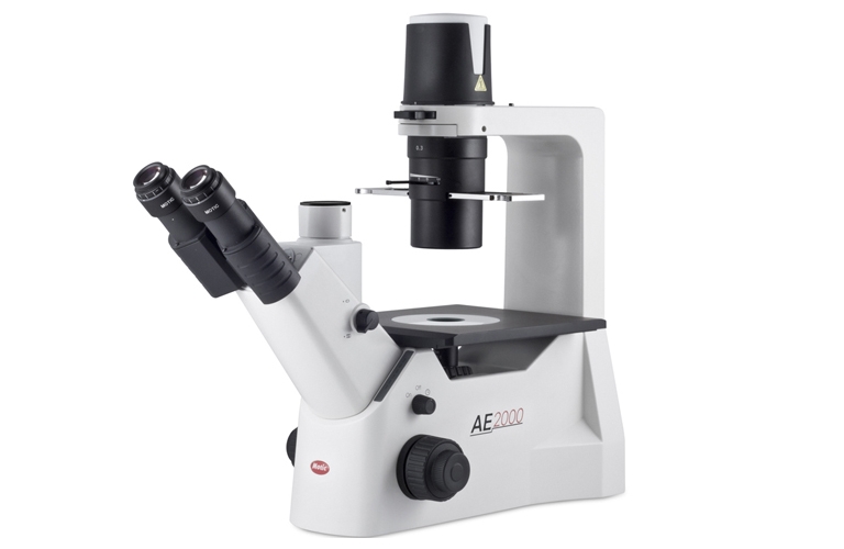
Motic AE 2000 |
|
|
|
Leica MZ6 dissecting stereomicroscope |
|
|
Brunel dissecting microscope |
|
|
 |
Zeiss stereomicroscope |
|
|
|
Motic SMZ-171 Series |
|


Fluorescence microscopes
| Make/Model | Features | ||
|---|---|---|---|
 |
Zeiss Axioplan epifluorescence microscope |
|
|
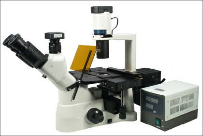 |
Brunel SP105F with pE300 LED |
|
|
Available Imaging Equipment - White City
Leica SP8 Thunder Widefield Microscope
Specifications
- Microscope
- DMi8 CS inverted
- XY scanning stage
- Z Galvo
- Universal insert
- Microplate insert
- LED illumination with DAPI, 470, RHOD and TXR filters
- Lasers
- 405nm
- 442nm
- Argon: 458nm, 476nm, 488nm, 496nm and 514nm
- DPSS: 561nm
- HeNe: 633nm
- Objectives
|
Correction
|
Magnification
|
NA
|
Immersion
|
Coverglass
|
Working distance
|
Other
|
|---|---|---|---|---|---|---|
| HC Plan APO | 10x | 0.40 | Dry | 0.17 | 2740um |
CS2 |
| HC Plan APO | 20x | 0.75 | Dry | 0.17 | 620um | CS2 |
| HC Plan APO | 40x | 1.30 | Oil | 0.17 | 240um | CS2 |
| HC Plan APO | 63x | 1.40 | Oil | 0.17 | 140um | CS2 |
- Detectors
- 2x PMT
- 3x HyD GaAsP
- Extras
- Lightning
- FRAP
- FRET
- 3D Visualiser
- Live Data Mode
Leica SP8 Lightning Confocal Microscope
Description
Confocal microscope without environmental control
Specifications
- Microscope
- DMi8 CS inverted
- XY scanning stage
- Z Galvo
- Universal insert
- Microplate insert
- LED illumination with DAPI, 470, RHOD and TXR filters
- Lasers
- 405nm
- 442nm
- Argon: 458nm, 476nm, 488nm, 496nm and 514nm
- DPSS: 561nm
- HeNe: 633nm
- Objectives
|
Correction
|
Magnification
|
NA
|
Immersion
|
Coverglass
|
Working distance
|
Other
|
|---|---|---|---|---|---|---|
| HC Plan APO | 10x | 0.40 | Dry | 0.17 | 2740um |
CS2 |
| HC Plan APO | 20x | 0.75 | Dry | 0.17 | 620um | CS2 |
| HC Plan APO | 40x | 1.30 | Oil | 0.17 | 240um | CS2 |
| HC Plan APO | 63x | 1.40 | Oil | 0.17 | 140um | CS2 |
- Detectors
- 2x PMT
- 3x HyD GaAsP
- Extras
- Lightning
- FRAP
- FRET
- 3D Visualiser
- Live Data Mode
Light and fluorescence microscopes
Specs coming soon
Zeiss MicroCT scanner
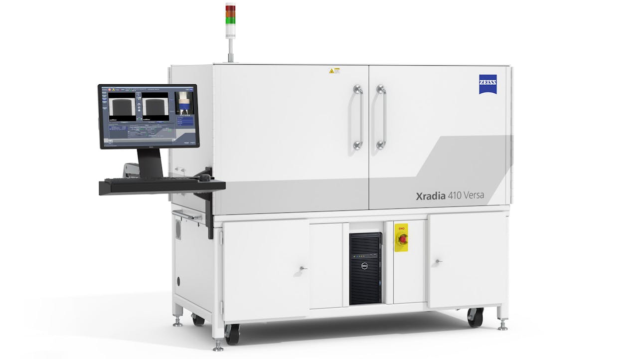
Imaging:
Spatial Resolution : 0.7 μm
Minimum Achievable Voxel : 70 nm
Resolution at a Distance (RaaD) at 50 mm working distance : 1 μm
X-ray source:
Type: Seal-transmission
Tube voltage range: 30 - 160 kV
Maximum output: 10 W
Detector system: ZEISS X-ray microscopes feature a detector turret with multiple objectives at different magnifications. Each objective features optimized scintillators that deliver the highest absorption contrast details.
Objectives: 0.4X, 4X and 20X
MicroscopyFAQ
Image analysis station

PC available for image analysis using Imaris and LASX
Specifications
- Imaris
- LASX
Links
Access protocol
Room safety induction and training required before access. Please request training in PPMS: https://ppms.eu/imperial/areq/?pf=5
Data Management
-- You are responsible for your data. --
Please move your data from the acquisition computers.
- They really aren't safe places to keep anything
- Please either move the data (rather than copy) or delete the local copies after you have them safely elsewhere
- You may keep a copy of your data on a microscope computer for up to one week while you are making sure you have it backed up to your own lab based storage location.
- We may delete any files without warning if space is required for experiments
- (Caveat for small files, we understand sometime it is useful to store a reference file (eg to reload settings) so it is fine to keep permanently a small volume of data in a clearly labeled folder)
- Any files on any computer should be in a folder with your full name or netid on the data drive (please don't use the desktop or "my documents" as it buries the files in places less easy to maintain.