Imperial College London

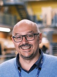
DrJorgeBernardino de la Serna

Faculty of MedicineNational Heart & Lung Institute

Senior Lecturer in Inhalation Toxicology and Pharmacology


//

Contact


+44 (0)20 7594 3277j.bernardino-de-la-serna Website


//

Location


CubicleSir Alexander Fleming BuildingSouth Kensington Campus

//

Summary


Publications

Citation

BibTex format

@article{Schneider:2018:10.1021/acs.nanolett.8b01190,
author = {Schneider, F and Waithe, D and Galiani, S and Bernardino, de la Serna J and Sezgin, E and Eggeling, C},
doi = {10.1021/acs.nanolett.8b01190},
journal = {Nano Letters},
pages = {4233--4240},
title = {Nanoscale spatiotemporal diffusion modes measured by simultaneous confocal and stimulated emission depletion nanoscopy imaging},
url = {http://dx.doi.org/10.1021/acs.nanolett.8b01190},
volume = {18},
year = {2018}
}

RIS format (EndNote, RefMan)

TY  - JOUR
AB - The diffusion dynamics in the cellular plasma membrane provide crucial insights into molecular interactions, organization, and bioactivity. Beam-scanning fluorescence correlation spectroscopy combined with super-resolution stimulated emission depletion nanoscopy (scanning STED–FCS) measures such dynamics with high spatial and temporal resolution. It reveals nanoscale diffusion characteristics by measuring the molecular diffusion in conventional confocal mode and super-resolved STED mode sequentially for each pixel along the scanned line. However, to directly link the spatial and the temporal information, a method that simultaneously measures the diffusion in confocal and STED modes is needed. Here, to overcome this problem, we establish an advanced STED–FCS measurement method, line interleaved excitation scanning STED–FCS (LIESS–FCS), that discloses the molecular diffusion modes at different spatial positions with a single measurement. It relies on fast beam-scanning along a line with alternating laser illumination that yields, for each pixel, the apparent diffusion coefficients for two different observation spot sizes (conventional confocal and super-resolved STED). We demonstrate the potential of the LIESS–FCS approach with simulations and experiments on lipid diffusion in model and live cell plasma membranes. We also apply LIESS–FCS to investigate the spatiotemporal organization of glycosylphosphatidylinositol-anchored proteins in the plasma membrane of live cells, which, interestingly, show multiple diffusion modes at different spatial positions.
AU - Schneider,F
AU - Waithe,D
AU - Galiani,S
AU - Bernardino,de la Serna J
AU - Sezgin,E
AU - Eggeling,C
DO - 10.1021/acs.nanolett.8b01190
EP - 4240
PY - 2018///
SN - 1530-6984
SP - 4233
TI - Nanoscale spatiotemporal diffusion modes measured by simultaneous confocal and stimulated emission depletion nanoscopy imaging
T2 - Nano Letters
UR - http://dx.doi.org/10.1021/acs.nanolett.8b01190
UR - http://gateway.webofknowledge.com/gateway/Gateway.cgi?GWVersion=2&SrcApp=PARTNER_APP&SrcAuth=LinksAMR&KeyUT=WOS:000439008300026&DestLinkType=FullRecord&DestApp=ALL_WOS&UsrCustomerID=1ba7043ffcc86c417c072aa74d649202
UR - http://hdl.handle.net/10044/1/69260
VL - 18
ER -