Group III - Collectins







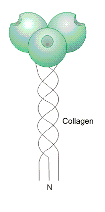 The collectin group of CTLD-containing secreted proteins has six members
in the mouse, but only five in humans, because the human gene for
mannose-binding protein (MBP)-A is a pseudogene. MBP-A and MBP-C are
serum proteins, whereas surfactant protein (SP)-A and SP-D are secreted by
epithelial cells and decorate mucosal surfaces, notably as
components of pulmonary surfactant. Little is known
about collectins K1 and L1. A collectin monomer has an N-terminal
collagen-like region, which is followed by a section of heptad repeats,
and a C-terminal CTLD. Collectin monomers assemble into characteristic
homotrimeric units, each of which has a collagen-like triple helical tail,
a coiled-coil neck region, and a rigid cluster of CTLDs. Multiple
homotrimeric units associate via disulphide bond formation to form
supramolecular complexes which are either sertiform/bouquet-like (MBP and
SP-A) or cruciform/cross-shaped (SP-D).
The collectin group of CTLD-containing secreted proteins has six members
in the mouse, but only five in humans, because the human gene for
mannose-binding protein (MBP)-A is a pseudogene. MBP-A and MBP-C are
serum proteins, whereas surfactant protein (SP)-A and SP-D are secreted by
epithelial cells and decorate mucosal surfaces, notably as
components of pulmonary surfactant. Little is known
about collectins K1 and L1. A collectin monomer has an N-terminal
collagen-like region, which is followed by a section of heptad repeats,
and a C-terminal CTLD. Collectin monomers assemble into characteristic
homotrimeric units, each of which has a collagen-like triple helical tail,
a coiled-coil neck region, and a rigid cluster of CTLDs. Multiple
homotrimeric units associate via disulphide bond formation to form
supramolecular complexes which are either sertiform/bouquet-like (MBP and
SP-A) or cruciform/cross-shaped (SP-D).
Carbohydrate recognition is central to the functions of MBP, SP-A and SP-D as pathogen-binding molecules of the innate immune system. MBP aids the elimination of the circulating microorganisms that it binds by initiating the lectin branch of the complement pathway and by stimulating phagocytosis. MBP mutations that reduce the efficiency of complement activation produce immunodeficiency in young children and immunocompromised adults. SP-A and SP-D engage microorganisms, including inhaled pathogens, to prevent them from adhering to epithelia, and to create aggregates that may be phagocytosed. These proteins also exert a direct antimicrobial effect by increasing the permeability of bacterial cell membranes. SP-A and SP-D interact with a range of leukocytes, and modulate the phagocytic activity of neutrophils and macrophages.
The CTLDs in MBP, SP-A and SP-D bind mannose-type sugars using a simple binding site which interacts only with terminal residues. However, collectin oligomers bind with high affinity and specificity to arrays of structurally complex and diverse glycans found on pathogen surfaces, due to the multivalent nature of the collectin complexes and the fixed geometry of the CTLDs.
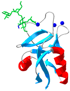
Structure of rat MBP CRD with a bound high mannose oligosaccharide
Mannose residues are shown in green, GlcNAc residues in blue, and Ca2+ ions in dark blue. Protein Data Bank structure ID: 2MSB.
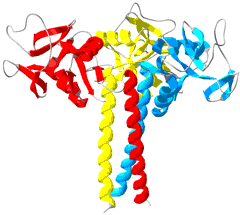
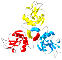
One polypeptide is shown in red, one in yellow, and one in blue. Protein Data Bank structure ID: 1RTM.


