Group IV - Selectins







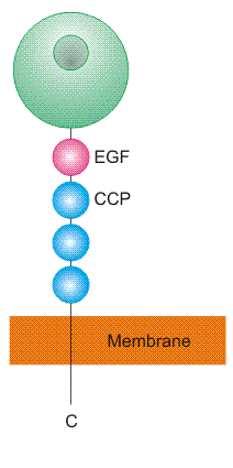 The selectin group of CTLD-containing cell surface proteins has
three members in both human and mouse. P-selectin is expressed on
endothelial cells and platelets, E-selectin on endothelial cells, and
L-selectin on leukocytes. Selectins are type 1 transmembrane
proteins, in which a single CTLD is projected from the cell surface by a
flexible neck consisting of an epidermal growth factor (EGF)-like domain
and a number of complement control repeats. The short cytoplasmic tail
influences subcellular localization and is involved in signalling.
The selectin group of CTLD-containing cell surface proteins has
three members in both human and mouse. P-selectin is expressed on
endothelial cells and platelets, E-selectin on endothelial cells, and
L-selectin on leukocytes. Selectins are type 1 transmembrane
proteins, in which a single CTLD is projected from the cell surface by a
flexible neck consisting of an epidermal growth factor (EGF)-like domain
and a number of complement control repeats. The short cytoplasmic tail
influences subcellular localization and is involved in signalling.
The selectins are leukocyte adhesion molecules. Weak, transient interactions between selectin CTLDs and specific glycan structures on cell surface glycoproteins bring about rolling of leukocytes on endothelia. Following changes in the cytoskeleton and in the expression of cell adhesion molecules (including internalization and degradation of the selectins themselves), these contacts are succeeded by integrin-mediated interactions that arrest leukocytes and permit leukocyte migration across endothelia. P- and E-selectin recruit leukocytes to sites of infection. Proinflammatory signals cause P-selectin to be transported to the endothelial cell surface from Weibel-Palade bodies and induce transcription of E-selectin. These selectins bind to leukocyte glycoproteins, principally P-selectin glycoprotein ligand (PSGL)-1 and E-selectin ligand (ESL)-1, initiating leukocyte migration into inflamed tissues. Platelet P-selectin is mobilized from storage in alpha-granules by activating stimuli, and mediates platelet-leukocyte adhesion in coagulation and thrombosis. L-selectin mediates lymphocyte homing to peripheral lymph nodes by binding to glycoprotein ligands on high endothelial venules, such as mucosal addressin cell adhesion molecule (MadCAM)-1 and CD34. L-selectin also directs leukocytes to sites of infection by binding to glycoproteins on inflamed endothelia. It mediates secondary tethering of leukocytes by binding to glycoproteins such as PSGL-1. In patients with type II leukocyte adhesion deficiency (LAD-II), the absence of fucosylated glycans precludes selectin-mediated leukocyte adhesion, increasing susceptibility to infection. The selectins are also signalling molecules: they initiate outside-in signalling upon ligation, they may be regulated by inside-out signalling through changes in oligomeric state and cytoskeletal association, and they may affect signalling by their interaction partners.
The CTLD in selectins is specific for sialyl-Lewisx and closely-related structures. Fucose is bound at the primary binding site, which is of the mannose-binding subtype, with sialic acid accommodated by a secondary binding site. In P-selectin a region of positive charge interacts with sulphated tyrosine residues in PSGL-1, and high affinity L-selectin ligands also exhibit sugar or tyrosine sulphation.
- Structure of human P-selectin
- bound to PSGL-1
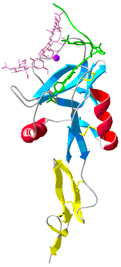
Shown in green is the backbone of PSGL-1 plus the side chains of two sulphated Tyr residues. Shown in pink is the PSGL-1 glycan. Protein Data Bank structure ID: 1G1S.


