This seminar has been postponed.
Abstract
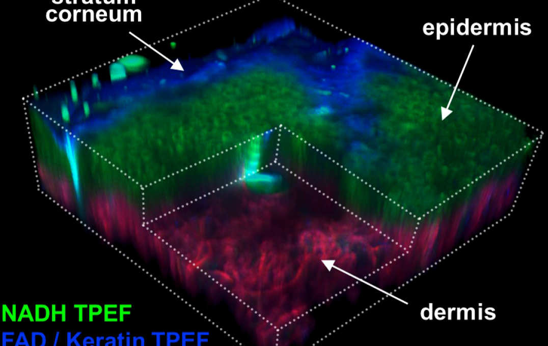
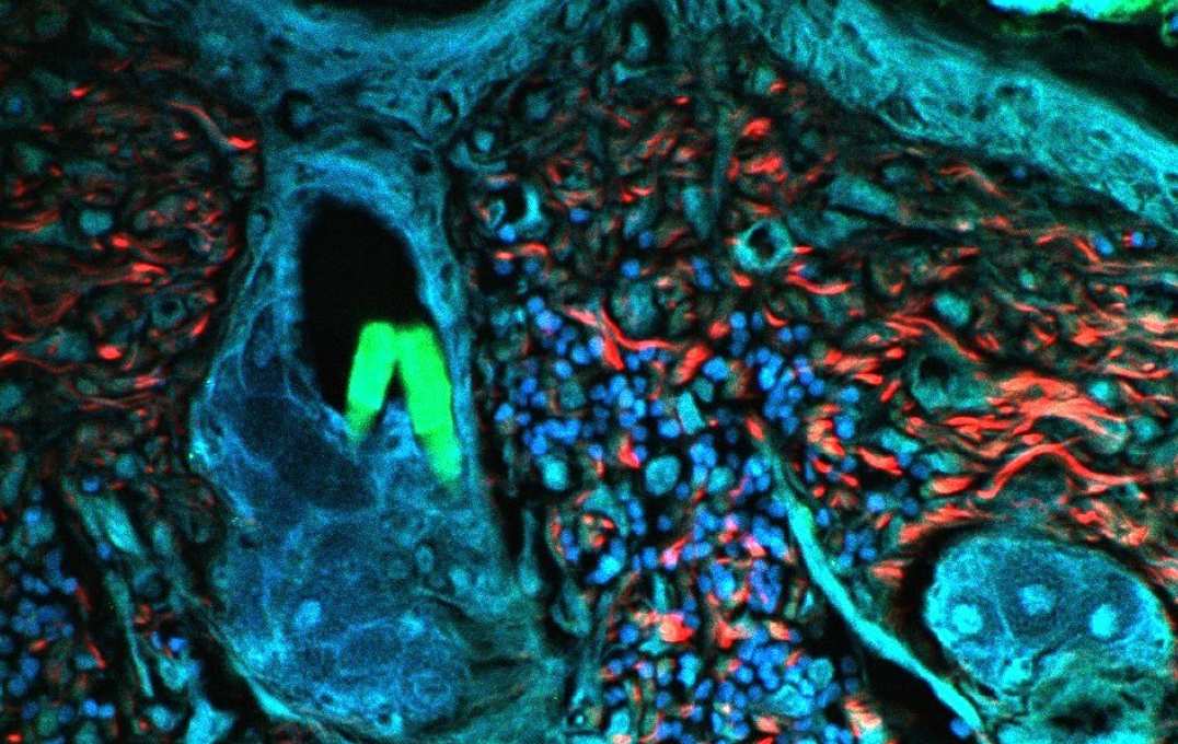
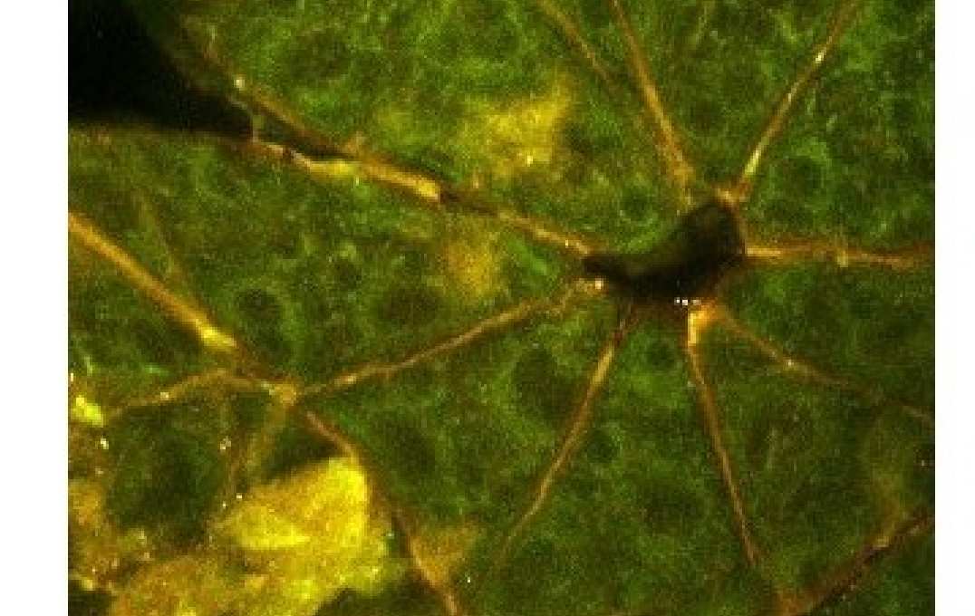
Non-healing wounds, such as diabetic foot ulcers, are challenging to diagnose and treat due to their numerous etiologies and the variable efficacy of wound care products. There is a critical need to develop new diagnostic technologies and quantitative biomarkers that are sensitive to specific wound characteristics. Multiphoton microscopy techniques are well-suited for 3D skin imaging and are capable of non-invasively detecting autofluorescence from metabolic cofactors (NADH and FAD) without the need for exogenous dyes. Using an optical redox ratio of FAD/(NADH+FAD) autofluorescence and fluorescence lifetime imaging, we demonstrate differences in skin wound metabolism between aged, diabetic, and young mice through longitudinal monitoring over 10 days post-wounding. These changes in keratinocyte metabolism are associated with the demands of proliferation and migration during re-epithelialization. Our work demonstrates the potential of label-free autofluorescence imaging to distinguish different underlying causes for delays in healing, which may be used in the future to provide patient-specific treatment strategies in the clinic.
Biography:
Kyle P. Quinn, PhD, is an assistant professor of biomedical engineering at the University of Arkansas. Dr. Quinn received his B.S. degree in Biomedical Engineering from the University of Wisconsin-Madison, and earned his Ph.D. in Bioengineering from the University of Pennsylvania. As a postdoctoral fellow at Tufts University, he was awarded an NIH Ruth L. Kirschstein National Research Service Award and an NIH Pathway to Independence Award (K99). Since joining the Department of Biomedical Engineering at the University of Arkansas, he has received funding through the NIH R00, R21, and R01 mechanisms, DoD STTR and CDMRP projects, and the NSF CAREER award. His overall research interests are in developing and utilizing non-invasive label-free multiphoton microscopy techniques to characterize the spatiotemporal patterns of disease progression and tissue repair processes. His research group is currently focused on using two-photon excited fluorescence, second harmonic generation, fluorescence lifetime imaging, and coherent anti-Stokes Raman spectroscopy to establish quantitative optical biomarkers for a variety of diseases and conditions.