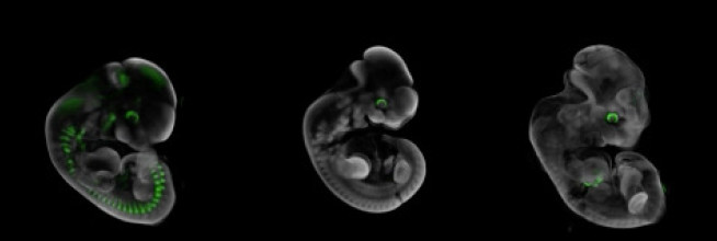
Cross-Faculty researchers have developed pioneering epigenetic imaging which allows in vivo imaging of epigenetic change using bioluminescence and photoacoustic imaging methods. The epigenetic imaging is used to study genes in X chromosome inactivation.
Through combining epigenetic expertise from Fisher and Sardini (Institute of Clinical Sciences, Medicine) and photonics expertise from French and McGinty (Department of Physics, Natural Sciences) alongside industrial collaboration with Perkin Elmer and Fujifilm VisualSonics. They have developed sensitive bioluminscence and photoacoustic methods to allow luciferase and LacZ reporter gene expression to be non-invasively imaged in mice in vivo and in utero.
Publications
Van de Pette, Mathew; Abbas, Allifia; Feytout, Amelie; Mcnamara, Gráinne; Bruno, Ludovica; K. To, Wilson; Dimond, Andrew; Sardini, Alessandro; Webster, Zoe; Mcginty, James; Paul, Eleanor; A. Ungless, Mark; M.W. French, Paul; J. Withers, Dominic; Uren, Anthony; C. Ferguson-Smith, Anne; Merkenschlager, Matthias; M. John, Rosalind; G. Fisher, Amanda. (2017). Visualizing Changes in Cdkn1c Expression Links Early-Life Adversity to Imprint Mis-regulation in Adults. Cell Reports. 18. 1090-1099. 10.1016/j.celrep.2017.01.010