Incredible images with dark Victorian past scoop top prize
by Kate Wighton
Inner core of Victorian bones
A set of startling images of the bones of Victorian children have won a prestigious international award.
The pictures, which were created by an undergraduate at Imperial College London, show in incredible detail the inner core of our bones, and provide a window into how bones grow in the womb, and develop in early years.
They have this week been named as one of twenty winning images in the Wellcome Image Awards, an international competition that showcases the best of biomedical imaging.
From these images we were able to see that the core of our bones starts to change when we start walking
– Dr Frank Acquaah
Award winner
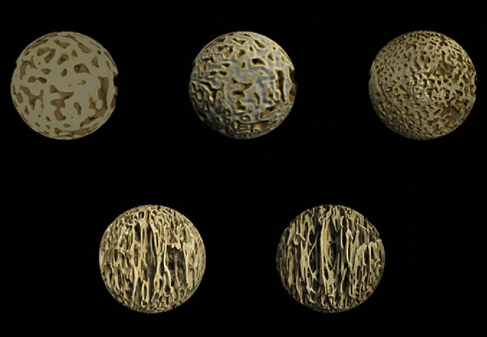
The images were created using samples of bone from Victorian children who died before birth or in infancy. The bones, which were preserved by the Royal College of Surgeons, were from a foetus at around 6 months, at 9 months, a child under the age of one, at just over one year old, and at two-and a-half years old.
The images, which were created by Dr Frank Acquaah while studying for a BSc in Surgery and Anaesthesia at Imperial, show the inner spongy layer of our bones called the trabecular layer. This layer, which is a type of lattice, lends bones their strength and is often found to be weakened in bone conditions such as osteoporosis.
To gain a better understanding of the bone conditions that strike children and adults, Dr Acquaah scanned the bone samples, which were taken from the spine, with a special type of scan called a micro-CT Scan. This is able to show structures in incredible detail. The images were then processed with a computer programme to create a 3D image.
Dr Acquaah, who created the images in the Musculoskeletal Lab at Imperial, explained: “We know the trabecular layer is crucial to bone strength, but studying it during development is very difficult. However, from these images we were able to see that the core of our bones starts to change when we start walking, which is around the age of two. Here it starts to develop so-called struts, in the same direction of the vertical force on the spinal cord."
: A foetus at 6 months, at 9 months, and a child between birth and one year. Bottom row (L-R): At just over one year old, and at two and a half years old.")
Images of the inner core of bones from the spine, across different ages. Top row (from L-R): A foetus at 6 months, at 9 months, and a child under the age of one. Bottom row (L-R): A child aged just over one year old, and at two and a half years old.
Dr Acquaah adds that although these samples are from children in the 1800s, previous work has shown the samples are similar enough to the bones of modern humans for the findings to be relevant today.
He adds: “It was such a surprise to receive the Wellcome Image award. I’m incredibly proud, and also very pleased that so many people will be able get a flavour of the fascinating work at the Musculoskeletal lab at Imperial.”
Dr Richard Abel, from the Department of Surgery and Cancer at Imperial, and who was Dr Acquaah’s supervisor during his studies adds: “In the Musculoskeletal lab we are trying to understand how we can promote healthy ageing. We are realising that health in old age is strongly influenced by our early development, even during gestation. With Frank’s images we are trying to capture the earliest changes in bone structure that occur around birth and when we stand up and walk. Early exercise, even kicking and punching in utero, may be important for developing strong bones.”
All the winning Wellcome images can be viewed here.
Article supporters
Article text (excluding photos or graphics) © Imperial College London.
Photos and graphics subject to third party copyright used with permission or © Imperial College London.
Reporter
Kate Wighton
Communications Division