Human brain cell transplant offers insights into neurological conditions
by Kate Wighton
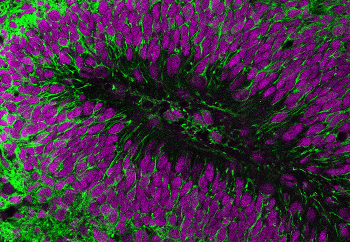
Transplanted human brain cells (green) and nuclei (purple)
Scientists have created a ‘window’ into the brain, which enables researchers to watch in detail how human brain cells connect to each other.
In the new study, led by scientists from Imperial College London in collaboration with a group from the University of Cambridge, researchers for the first time successfully transplanted human brain cells into a mouse brain, and watched how they grew and connected to each other. This allowed the team to study the way human brain cells interact in a more natural environment than previously possible.
We were hoping a few of the human brain cells would grow within the mouse brain – but we were stunned to see the human brain cells thrive, and soon talk and work together Dr Vincenzo De Paola Study author
The team, funded by the Medical Research Council, used the technique to model Down's syndrome, using cells donated by two individuals with the condition.
The scientists say the technique could be used to study a range of brain conditions in the future, including schizophrenia, dementia or autism.
The study, published today in the journal Science, describes how researchers saw differences in the brain cells from the individuals with Down's syndrome compared to brain cells from a person without the condition.
Although some of the connections formed between the brain cells from the individuals with Down's syndrome were more stable and abundant, they communicated in a slightly less coordinated fashion.
Dr Vincenzo De Paola, lead author of the research from Imperial’s Institute of Clinical Sciences, said: “It’s been a fantastic team effort and I’m grateful to the many scientists who participated in this study, as well as to the people who donated tissue samples for this research.Our results suggest the reduced coordinated activity and increased stability of connections in Down's syndrome may be linked to cognitive function. Figuring this out would be an important piece of the puzzle, and we hope to have an answer soon.”
Connections seen for first time
Professor Rick Livesey, ioint co-corresponding author from the University of Cambridge’s Wellcome/Cancer Research UK Gurdon Institute, commented: “Working together with the Imperial team has allowed us to extend our previous work on making stem cells and nerve cells from people with Down's syndrome, to study how those nerve cells develop and function when put in a living brain. We are very excited by how much we have learned and the new avenues this has opened up for understanding Down's syndrome”.

Transplanted human brain cells (green) and nuclei (purple).

Synaptic connections (green) in human brain cell transplant (purple)

Blood supply (purple) in transplanted human brain cells (green)
Dr Raquel Real, a neurologist from Dr De Paola’s group at Imperial College London and joint first author of this study, added: “The transplantation of human brain cells has allowed us to monitor their maturation over time. Ultimately, we detected that cells from Down syndrome individuals are not as active as normal cells at a crucial stage in their development, and this could have important implications for some of the symptoms of this condition”.
Dr De Paola added: “Scientists have been struggling to develop a way of monitoring live human cells and their connections in the brain. This new approach may have taken us one step closer to this.”
Crucially, the technique allows scientists to study how brain cells communicate, explained Dr De Paola: “The connections between brain cells, which enable them to talk to each other, is often the first thing to be damaged in conditions such as dementia and Parkinson’s. This happens long before the brain cells themselves start to die. But the connections are so tiny, that no type of scanning tool we currently use, such as MRI or PET scans, can see them. But the revolutionary microscopy technique used in this study – called in vivo 2-photon microscopy – allows us to not only see individual live brain cells, but also the connections between them.”
Growing brain cells from skin cells
In the study, Dr Manuel Peter from the Gurdon Institute created human brain cells by reverse-engineering skin cells. This process involved taking a few skin cells from volunteers with Down's syndrome, and then reprogramming them in the lab to form brain cells. They then engineered those neurons so that their activity could be monitored in the live mouse brain, using 2-photon microscopy.
Joint first authors Dr Raquel Real and Dr Antonio Trabalza from the Imperial College London group were then able to implant these human neurons in the brain of live mice.
Dr De Paola explained: “The human brain cells not only formed complex networks, but also started communicating in a way that was very similar to normal brain cells. We were hoping a few of the human brain cells would grow within the mouse brain – but we were stunned to see the human brain cells thrive, and soon talk and work together. Eventually the human cells constituted a large area in the mouse brain.”
However, he cautioned: “It is still not clear to what extent the transplanted human brain cells resemble the organisation and complexity of their counterparts in the human brain. We now need to investigate this with further experiments.”
The team now hope to refine this technique, and potentially use this approach to study other neurological conditions.
The work also received support from the the Rosetrees Trust, the Wellcome Trust and Alzheimer’s Research UK
-
"In vivo modeling of human neuron dynamics and Down syndrome" by R. Real at el. is published in Science
Article supporters
Article text (excluding photos or graphics) © Imperial College London.
Photos and graphics subject to third party copyright used with permission or © Imperial College London.
Reporter
Kate Wighton
Communications Division