We have developed a range of optical tomography instruments for preclinical imaging of disease models or 3D imaging of tissue samples for pathology. The tremendous advances in chemical clearing techniques to render tissue samples transparent has opened up 3D histopathology of intact tissue samples and OPT is well-suited to this application. For in vivo preclinical imaging, OPT may be applied to small, semi-transparent organisms such as nematodes and zebrafish. To widen access to this capability, we developed OPTImaL, a cost effective open hardware implementation of OPT with associated open-source software. For optical in vivo imaging of mammalian disease models, we have explored diffuse optical tomography incorporating time-gated imaging.
Optical Tomography
For basic 3D imaging of transparent samples, we have developed a low-cost open configuration (Dual-OPT) for OPT that provides imaging at x4 and unity magnification and for fluorescence OPT can utilise multiple LED excitation sources or a laser-based source with a suitable dynamic diffuser to provide reasonably uniform illumination intensity for excitation. We have implemented OPT with a fibre-laser-pumped supercontinuum source or with diode lasers and have applied such instruments to a wide range of cleared samples, including cleared murine pancreas samples for studies of diabetes [1]. We also implement simple transmission OPT using an LED backlight to provide extended
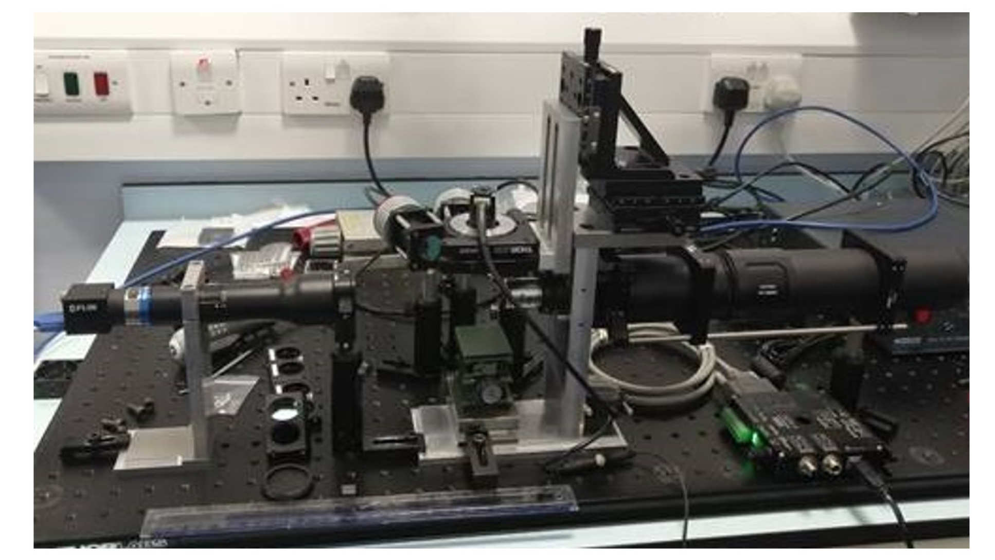
openScopes OPT system with imaging arms for x4 and x1 magnification and LED excitation
For researchers wishing to implement OPT of smaller (< 1mm diameter) samples at minimal cost, we have also developed a simple OPT module that can be added to a standard inverted fluorescence. This OPTiM approach is based on an OPT microscope plate adaptor module [2] makes use of the existing instrument components, including camera, objective lenses and light sources.
Since fluorescence OPT entails the acquisition of wide-field (projection) images, it can be easily adapted to FLIM OPT, e.g. using a GOI for time-gated imaging. We demonstrated this for fixed cleared or transparent samples [3] and progressed to in vivo imaging of naturally transparent live zebrafish embryos expressing fluorescent proteins [4], including rapid 3D FLIM of zebrafish embryos expressing a FRET biosensor for caspase 3 as a reporter of apoptosis [5].
For OPT of live samples, such as zebrafish, it is important to minimise the light dose and the image acquisition time since the samples must be maintained under anaesthesia. It is therefore desirable to minimise the number of acquired projection images – a particularly important consideration for OPT FLIM where sequences of time-gated images are acquired at each projection angle. However, to realise “lossless” (i.e. full Shannon-Nyquist sampling) 3D imaging, the number of projection images required is proportional to the number of resolution elements in the projection image and is typically several hundred. Acquiring fewer projection images can result in “streak” artefacts in the final images reconstructed using the standard filtered back projection (FBP) algorithm.
Since considerations of phototoxicity and experimental convenience usually result in undersampled OPT data sets, we have worked to mitigate the streak artefacts. In one approach we incorporate a measured modulation transfer function (MTF) in the reconstruction process [6], which improves the image quality. For in vivo imaging of live zebrafish – and particularly for longitudinal studies of adult zebrafish – we are constrained to total acquisition times of a few minutes and, when imaging relatively sparsely expressed fluorescent proteins, need to reconstruct 3D images from much less than 100 projection images. For this, we utilised compressive sensing techniques and, working with colleagues from UCL, demonstrated that we could reconstruct useful 3D images from fewer than 40 projection images using an iterative algorithm [7]. While this worked well, the iterative reconstruction process was quite slow, and we showed that this could be accelerated using a convolutional neural network (CNN) with FBP to reconstruct the same undersampled OPT data sets with improved speed and image quality compared the iterative compressed sensing algorithm [8].
We have also worked to enhance the imaging performance of OPT in terms of spatial resolution. In general, there is a trade-off between sample size and achievable spatial resolution. This arises from the assumption of parallel rays through the sample that is required for 3D image reconstruction using FBP. For OPT, the image can be reconstructed from ~parallel rays extending through half the sample (noting that it is imaged from both sides during a full scan and any signal from strongly diverging rays does not contribute to the reconstructed image). Thus, the sample radius sets the minimum depth of field of the optical imaging system, which in turn sets the maximum numerical aperture and therefore the achievable spatial resolution. This limit can be overcome using focal scanning where a higher NA lens is scanned such that its reduced depth of field is translated throughout the (first half of) the sample. The out-of-focus light is detected and reduces the dynamic range of the projection images but does not contribute to the FBP-reconstructed images. We implemented a cost-effective focal scanning OPT system using an electrically tunable liquid lens [9].
An alternative approach to increase the spatial resolution is to use multiple optical imaging systems of higher NA with each being focussed to provide reconstructed 3D images from different annular slices of the sample. The use of multiple imaging systems increases the light collection efficiency as we demonstrated in a 2-channel instrument [10]. Subsequently we implemented this approach for longitudinal imaging of a live adult zebrafish cancer model to study progression of angiogenesis and tumour development [11].
This multiplexed OPT approach can also be used to implement simultaneous multispectral imaging if two similar optical systems are focussed to the sample region or it can be used for 3D cell tracking where two imaging systems focussed on the same sample region at different projection angles can be used to determine the 3D location of features at the camera frame rate using triangulation while the sample is rotated to also permit reconstruction of the OPT image. We implemented this to track the 3D trajectories of neutrophils in a live zebrafish embryo [12] as a tool to study responses to inflammation.
When imaging juvenile or adult zebrafish, the image quality is typically reduced by light scattering and this will degrade OPT fluorescence images. To reduce the impact of scattering, we implemented a parallelised form of line-illumination and confocal detection in each projection image and then scanned the lines to acquire complete projection images with improved spatial resolution compared to standard wide-field projection images. We applied this approach we describe as “slice OPT” [13] to image live juvenile zebrafish and demonstrated an improvement in image quality. This slice-OPT technique will only work while there is a useful unscattered (ballistic) light component in the detected signal illumination.
When applying optical tomography to highly scattering samples, where the light can be considered to be isotropic, it is possible to reconstruct 3D images using the diffusion approximation. We have worked with colleagues at UCL to develop time-resolved diffuse fluorescence tomography (DFT) with the goal to undertake FLIM FRET based studies in live disease models. Since time resolved detection can be used to improve the image resolution of DFT, it is synergistic to combine DFT with FLIM/FRET. We focussed on DFT using cells expressing fluorescence proteins, demonstrating this for the first time [14] and progressing to demonstrate DFT FLIM FRET of cells embedded in a scattering phantom [15]. We then implemented in vivo FLIM FRET in a mouse model expressing a GFP/mCherryFP based FRET construct [16]. The use of visible fluorescence proteins limited the tissue depth from which we could obtain a useful signal and we believe that future efforts in this direction should be undertaken with NIR emitting fluorescent biosensors.
Optical tomography references
- Ablation of AMP-activated protein kinase alpha 1 and alpha 2 from mouse pancreatic beta cells and RIP2.Cre neurons suppresses insulin release in vivo,
G. Sun, A. I. Tarasov, J. McGinty, A. McDonald, G. D. Xavier, T. Gorman, A. Marley, P. M. French, H. Parker, F. Gribble, F. Reimann, O. Prendiville, R. Carzaniga, B. Viollet, I. Leclerc, and G. A. Rutter,
Diabetologia 53 (2010) 924-936 - OPTiM: Optical projection tomography integrated microscope using open-source hardware and software.
Watson T, Andrews N, Davis S, Bugeon L, Dallman MD, McGinty J
PLoS ONE 12 (2017) e0180309. - Fluorescence Lifetime Optical Projection Tomography
J. McGinty, K. B. Tahir, R. Laine, C. B. Talbot, C. Dunsby, M. A. A. Neil, L. Quintana Rio, J. Swoger, J. Sharp and P. M. W. French
J Biophotonics 1 (2008) 390-394 - In vivo fluorescence lifetime optical projection tomography
J. McGinty, H. B. Taylor, L. Chen, L. Bugeon, J. R. Lamb, M. J. Dallman, and Paul M. W. French
Biomed. Opt. Expr. 2 (2011) 1340-1350 - Visualising apoptosis in live zebrafish using fluorescence lifetime imaging with optical projection tomography to map FRET biosensor activity in space and time
N. Andrews, M-C Ramel, S. Kumar, Y. Alexandrov, D. J. Kelly, S. C. Warren, L. Kerry, N. Lockwood, A. Frolov, P. Frankel, L. Bugeon, J. McGinty, M. J. Dallman* and P. M. W. French*
J. Biophoton, 9 (2016) 414–424. - Incorporation of an experimentally determined MTF for spatial frequency filtering and deconvolution during optical projection tomography reconstruction,
L. Chen, J. McGinty, H. B. Taylor, L. Bugeon, J. R. Lamb, M. J. Dallman, and P. M. W. French
Optics Express, 20, (2012), 7323-7337 - Accelerated Optical Projection Tomography Applied to In Vivo Imaging of Zebrafish,
T. Correia 1, N. Lockwood, S. Kumar, J. Yin, M-C Ramel, N. Andrews, M. Katan, L. Bugeon, M J Dallman, J McGinty*, P. Frankel*, P. M W French* and S. Arridge*
PLoS ONE 10(2015): e0136213 - Convolutional neural networks for reconstruction of undersampled optical projection tomography data applied to in vivo imaging of zebrafish,
Samuel P. X. Davis, Sunil Kumar, Yuriy Alexandrov, Ajay Bhargava, Gabriela da Silva Xavier, Guy A. Rutter, Paul Frankel, Erik Sahai, Seth Flaxman, Paul M. W. French* and James McGinty*
J Biophotonics, 112 (2019) e201900128 - Remote focal scanning optical projection tomography with an electrically tunable lens,
L. Chen, S. Kumar, D. Kelly, N. Andrews, M. J. Dallman, P. M. W. French, J. McGinty,
Biomed. Opt. Exp. 5(10): 3367-3375 (2014). - Simultaneous angular multiplexing optical projection tomography at shifted focal planes
L. Chen. J. McGinty and P. M. W. French
Opt Lett 38, (2013) 851 - Quantitative in vivo optical tomography of cancer progression & vasculature development in adult zebrafish,
S. Kumar, N. Lockwood, M-C. Ramel, T. Correia, M. Ellis, Y. Alexandrov, N. Andrews, R. Patel, L. Bugeon, M. J. Dallman, S. Brandner, S. Arridge, M. Katan, J. McGinty,*, P. Frankel,*, P. M. W. French*
Oncotarget.7 (2016) 43939-43948 - Mesoscopic in vivo 3-D tracking of sparse cell populations using angular multiplexed optical projection tomography, L Chen, Yuriy A, Sunil Kumar, N Andrews, M J. Dallman, P M. W. French* and J McGinty*, Biomedical Optics Express 6 (2015) 1253- 1261
- Slice-illuminated optical projection tomography
S. P. X. Davis, L. Wisniewski, S. Kumar, T. Correia, S. R. Arridge, P. Frankel, J. McGinty, and P. M. W. French
Optics Letters 43 (2018) 5555-5558 - Fluorescence lifetime tomography of live cells expressing EGFP embedded in a scattering medium exhibiting background autofluorescence,
V. Y. Soloviev, J. McGinty, K. B. Tahir, M. A. A. Neil, A. Sardini, J. V. Hajnal, S. R. Arridge, and P. M. W. French
Opt. Lett. 32 (2007) 2034-2036 - 3-D imaging of Förster resonance energy transfer in turbid media by tomographic fluorescence lifetime imaging
J. McGinty, V. Y. Soloviev, K. B. Tahir,R. Laine,D. W. Stuckey, Joseph V. Hajnal,A. Sardini, P M.W. French, and S. R. Arridge,
Opt. Lett. 34 (2009) 2272-4 - In vivo fluorescence lifetime tomography of a FRET probe expressed in mouse
J. McGinty, D. W. Stuckey, V. Y. Soloviev, R. Laine1, M. Wylezinska-Arridge, D. J. Wells, S. R. Arridge, P. M. W. French, J. V. Hajnal and A. Sardini
Biomed. Opt. Expr. 2 (2011) 1907-1917