Leukaemia scientist Dr Cristina Lo Celso given Royal Microscopical Society award
Dr Lo Celso is awarded the RMS Medal for Life Sciences for ‘outstanding scientific achievements applying microscopy in the field of cell biology’.
The series of medals from the Royal Microscopical Society (RMS) is designed to recognise and celebrate individuals who make outstanding contributions to the field of microscopy across both the life and physical sciences.
In its citation, the RMS said Dr Cristina Lo Celso “has made paradigm-shifting contributions to the understanding of the dynamic cellular processes regulating haematopoietic stem cells in the bone marrow through the pioneering use of intravital microscopy.
Cristina’s achievements in applying microscopy to live science as demonstrated by publications, mentorship and scientific citizenship is outstanding. Royal Microscopical Society
“Cristina has demonstrated the rare ability to successfully tackle widely recognised technical challenges to advance the field of stem cell biology using advanced microscopy as well as novel image analysis and mathematical modelling.”
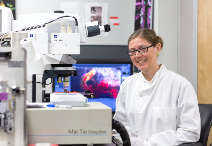
Dr Lo Celso investigates fundamental questions in stem cell biology, such as how a tissue regenerates over time and maintains organ functionality throughout a lifetime, and has been focusing on the haematopoietic (blood-cell forming) system as her experimental model.
Traditional techniques cannot reveal the dynamics of the blood stem cells and their interaction with stem cell niches, and these cells are inaccessible to direct observation because they reside in the bone marrow, deeply encased by bone.
While working at the Harvard Stem Cell Institute, Dr Lo Celso overcame this challenge using a combination of confocal and two-photon intravital microscopy of living mouse skull bone marrow, allowing her to visualize for the first time highly dynamic blood stem cell behaviours.
After starting her independent research group at Imperial College London in late 2009, she continued this approach and was instrumental in setting up intravital microscopy in the Facility for Imaging by Light Microscopy (FILM).
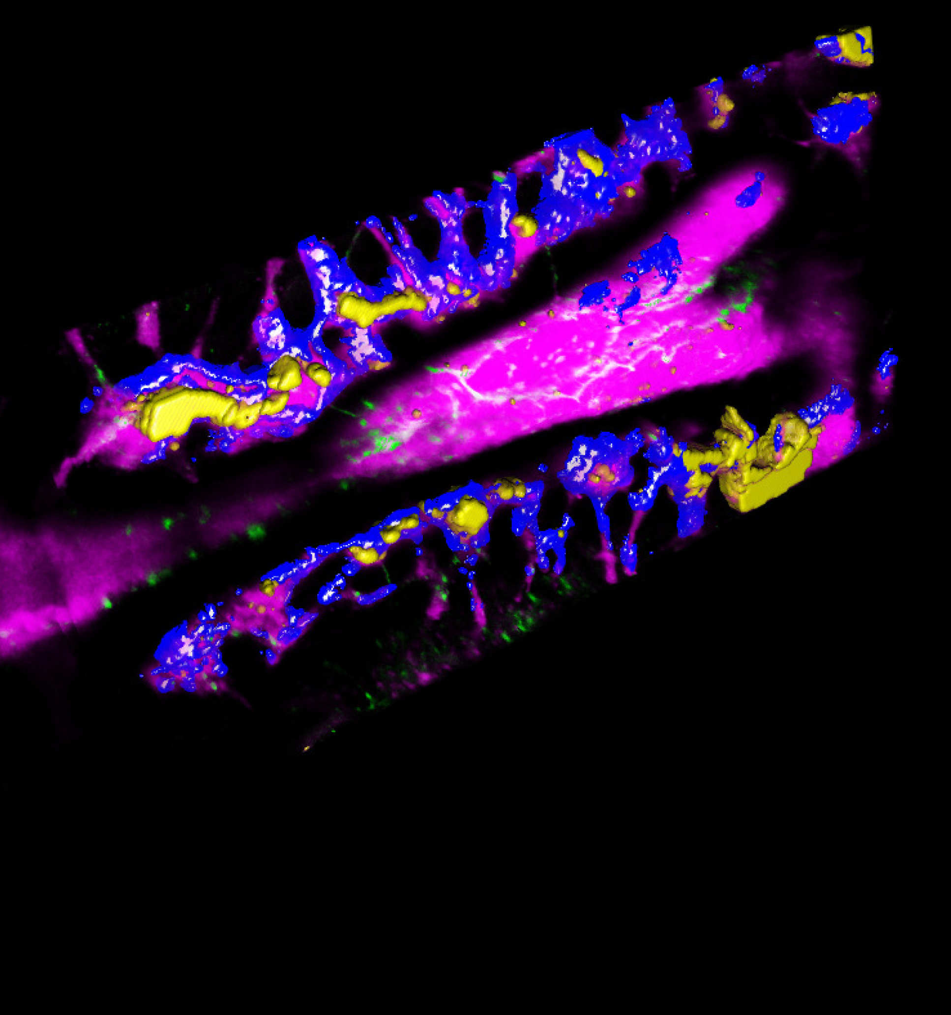
Since then she has achieved a number of ground-breaking discoveries, from uncovering changes in the behaviour of blood stem cells during steady state versus in response to natural infections, to achieving systematic quantification of stem cell localization relative to other bone marrow components.
Most recently she has found the unexpected migratory behaviour of leukaemia cells throughout disease development. This work shows early bone marrow infiltration of leukaemia cells in response to chemotherapy and their ability to rapidly destroy the niches that would normally support healthy blood production.
Dr Lo Celso adapted the imaging protocol to follow the same bone marrow areas over multiple days using implanted imaging windows. This work has deep implications for future development of improved leukaemia treatments, as she has also shown that her discoveries using mouse models hold true for human disease.
RMS said: “Cristina’s achievements in applying microscopy to live science as demonstrated by publications, mentorship and scientific citizenship is outstanding.”
Article text (excluding photos or graphics) © Imperial College London.
Photos and graphics subject to third party copyright used with permission or © Imperial College London.
Reporter
Hayley Dunning
Communications Division