Molecular insights into key drug target could help to enhance cancer treatments
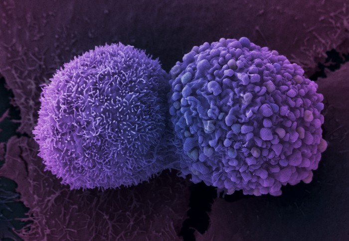
Lung cancer cells
New research reveals how a key protein in cells could be tweaked to make cancer treatments more effective.
Radiation and chemotherapy are designed to kill cancer cells. However, for many patients, cancer cells can survive even after being hit with high doses. To make these treatments more effective, scientists are focusing on ways to tweak the inner machinery of cancer cells to make them more susceptible to dying.
A team of scientists at Imperial College London and Washington University School of Medicine in St. Louis is making headway in such efforts. In a new study published in Nature Structural & Molecular Biology, the researchers have identified how a key protein in cancer cells changes shape to kick-start the repair of DNA damage caused by chemotherapy or radiation. Blocking this built-in repair mechanism with a drug has the potential to make chemotherapy or radiation more effective, according to scientists.
As this protein is essentially the same in lower organisms as it is in people, the researchers studied the version of the protein found in yeast, called Mec1. Mec1 and its human counterpart, ATR, are switched on when cells are stressed. These proteins are responsible for sensing and repairing DNA damage before cells replicate to prevent that damage from being passed on to daughter cells.
Normally, this activation is good, protecting healthy cells from DNA damage that could lead to cancer. However, in other cases, such as cancer therapy, doctors would like to turn off these repair mechanisms to make cancer cells more susceptible to death by further DNA damage. In this way, cancer cells – hit with radiation and chemotherapy – can be destroyed more easily.

The video shows two Mec1 molecules bound together (one is in colour on the left; the other is grey on the right). The side in colour shows how the protein moves to switch between active and inactive states. Credit: Luke Yates.
To understand how this protein functions, the Burgers Lab at the Washington University School of Medicine in St. Louis studied yeast with various mutations in this key protein. They discovered one mutant that forced the protein to be permanently switched on, but to identify how this was achieved, researchers needed to understand the molecular details.
Using high-resolution electron microscopes and cutting-edge detectors at the Electron BioImaging Centre and the Francis Crick Institute, researchers in the Zhang Lab at Imperial used the cryo electron microscopy technique to obtain high-resolution structures of both the native and mutant forms of the protein.
The molecular structures of both the inactive and active forms of this protein provide new insights into how changes from inactive to active take place, and how this protein is switched on to perform its vital functions. ATR kinase inhibitors are promising drugs for cancer treatment and the molecular structures provide the framework for improving current ATR kinase inhibitors or designing new ones.
Senior author Professor Xiaodong Zhang commented: “Cryo electron microscopy, a technique that won the 2017 Chemistry Nobel Prize, made it possible for us to visualize such complex large macromolecular machines in atomic details. Structures not only provide a molecular basis for how macromolecular machines function but are also required to explain how mutations cause disease, which will help with tailored treatment."
“We are grateful to the College, the generous funding from the Wellcome Trust, and our partners in London, which together have helped to establish the required cutting-edge infrastructure and made this type of work possible”.
Dr Luke A. Yates, a senior researcher in the Zhang lab and co-lead author, said: “Solving these large protein structures using cryogenic electron microscopy to such high-resolution has been a real career highlight (so far)."
"What was really wonderful to observe was how this relatively large protein twisted to place a small number of chemical groups (amino acid residues) into the correct position to allow it to perform its job. What is also quite neat is that these amino acid residues are held out of alignment to keep the protein turned off.”
Tannous EA, Yates LA, Zhang X, Burgers PM. Mechanism of auto-inhibition and activation of Mec1-ATR checkpoint kinase. Nature Structural & Molecular Biology. Nov. 9, 2020. DOI 10.1038/s41594-020-00522-0
This article was adapted from a press release by Washington University School of Medicine in St. Louis
Image credit: Anne Weston, Francis Crick Institute
Article supporters
Article text (excluding photos or graphics) © Imperial College London.
Photos and graphics subject to third party copyright used with permission or © Imperial College London.
Reporter
Ms Genevieve Timmins
Academic Services