
Bridging the gap between fundamental biophysics and medical research into the mechanisms underlying human diseases
The Section of Structural and Synthetic Biology aims to bridge the gap between fundamental biophysics and medical research into the mechanisms underlying human diseases. We are sited in South Kensington, as part of the Department of Infectious Disease at Imperial College London.
The Section of Structural and Synthetic Biology was set up in order to integrate fundamental structural and molecular biology directly with medical research at Imperial College London. Professor Harry Low heads up the section, which also includes the groups of Professor Paul Freemont, Professor Xiaodong Zhang, Professor Dale Wigley, Dr David Riglar , Dr Ravanish Krishna Kumar, Dr Szymon Manka and Dr Srinivasan Rengachari at present, with more scientists on the way.
Our research focuses on understanding the molecular mechanisms underlying factors linked to human disease, using a multi-disciplinary approach to dissect their structure and function in parallel. The interests of the investigators within the section span many fundamental cellular processes including chromatin remodelling, DNA replication, transcription, the repair of DNA damage, membrane fusion, protein degradation, signal transduction, and host-microbe interactions within the microbiota, all of which are linked to important human diseases. Our aim is to make the most direct contributions possible to understanding these conditions with the benefit of modern techniques.
The Section is an exciting and rewarding place to work, providing a hub of high-level knowledge bearing on the fundamentals of biochemistry, synthetic biology and structural biology, right in the heart of one of the world’s most important and vibrant cities. We are happy to hear from any researchers interested in asking about our work or thinking about coming to join us and to share our techniques and expertise with collaborators both in the UK and abroad.
Join us
The Section of Structural and Synthetic Biology at Imperial College is actively seeking to support candidates looking to lead their own laboratory through independent fellowship schemes. We welcome applications from scientists performing research using structural and/or synthetic and engineering biology in their many forms. Where relevant, joint applications with other sections in the Department of Infectious Disease may also be supported. We particularly encourage female scientists and those with diverse backgrounds to apply. The deadline to apply is Friday 12th September 2025.
About us
- Molecular mechanism - key to clinical progress
- Mind the gap - a multi-disciplinary approach to medical research
- Help us make a difference - collaboration and expansion
 Since the key molecules of life, DNA, RNA and protein, were understood in the 1950s, the molecular mechanisms underlying almost every aspect of biology have started to become incrementally clearer and clearer. We now have catalogues and blueprints, such as proteomes and genome sequences, for not just humans, but many of the most important disease-causing bacteria and viruses. Interpreting this wealth of data remains a fraught process however, the exact way in which molecules mesh to form the machinery of the cell requires their visualisation in atomic detail, which is still a difficult and costly endeavour. This process has had new life breathed into it over the past few years through great strides forward in the technique of electron cryo-microscopy, for which the Nobel prize for Chemistry was awarded to Dubochet, Frank and Henderson in 2017. Scientists within the Section have been intimately involved in the process of understanding proteins involved in immunology, cancer, and DNA damage repair through such novel approaches. Given the efforts required to eke such knowledge out of the molecules of life themselves, and the great potential for new treatments, new drugs or new approaches entirely, we are keen to share our work as widely and with as much impact as possible.
 The Section for Structural and Synthetic Biology was founded by Professors Paul Freemont and Xiaodong Zhang, both of whom have spent their careers committed to producing scientific results that have real-world applications. Having both seen many important mechanistic results founder in the gap between the laboratory bench and bedside, they reasoned that in order to continue to have a real impact through medical research, they would need to integrate directly into a medical centre of excellence. Imperial College is at the forefront of clinical research in the UK, and they, therefore, established the Section within what was formerly known as the Department of Medicine (now the Department of Infectious Disease) at Imperial.
The focus of work within the Section for Structural and Synthetic Biology is to build an understanding of disease mechanisms from the mechanistic level, looking at individual molecules, to cellular models and eventually clinical applications. This commitment has not only been to approach diseases of the Western World, as scientists within the section are also directly pursuing collaborations aiming to aid developing nations. Paul and Xiaodong are recruiting like-minded scientists, with impressive research track records and a similar shared interest in human disease mechanisms, expanding to tackle new problems.
 The scientists overseeing the Section are constantly looking for opportunities to expand both our collaborative network and our team here. We are on the lookout for like-minded scientists who wish to make a difference, and who are at any stage in their careers. In particular, scientists with a clinical or pharmacological bent would be welcomed with open arms; the Section remains a work in progress, and our commitment to promote collaboration and integration between structural and mechanistic understanding and the cellular and clinical aspects of medicine cannot make progress without active, prolonged engagement from both sides.
Research areas
In order to understand the molecular bases of human disease we are attempting to determine the molecular structures of the key complexes involved in the signaling process. Protein structure determination remains an extremely difficult process, frequently taking many years, however great strides forward have been made recently. To do this we are using both x-ray crystallography and electron cryo-microscopy; techniques that have each independently resulted in the award of the Nobel Prize for their application to structural biology.
Electron cryo-microscopy
Electron cryo-microscopy is the technique of the hour, it’s inventors, Richard Henderson, Jaques Debouchet and Joachim Frank, having been awarded the Nobel Prize for Chemistry in 2017. It is the latest and most powerful method to look at molecules in atomic detail. Whereas it was once derided as “blobology”, it is now frequently capable of reaching resolutions greater than 3 Å.
Samples of a molecule are frozen in vitreous (non-crystalline) ice, by plunging them into liquid ethane or propane. This preserves them immobile and protected, but in an almost native state. Once frozen, we subject the sample to a high-voltage (and therefore high-frequency) electron beam. Electrons are scattered by the sample, and because electromagnets can be used as lenses for them, we can recombine the scattered beam in order to generate phase-contrast, thereby retrieving the phase (in contrast to x-ray crystallography), and visualising the single particles in our sample.
Each independent image of a particle corresponds to a projection of its electron scattering potential from a single direction, and these individual images can be slotted back together in three dimensions to give the original scattering volume if we can work out the angle at which the particle was sitting in the ice. Because electrons also damage the sample, only a few electrons can be used before the particles start to become radiation damaged, and these images are also very noisy. It typically requires between tens of thousands and millions of particle images to determine the molecular structure of a sample.
Imperial has several electron microscopes available through the centre for structural biology, and is the founder member of the LonCEM consortium, tasked with siting an FEI Krios TEM at the Crick institute.
X-ray Crystallography
X-ray crystallography has long been the workhorse of the structural biologist. It was responsible for the first molecular structures of proteins in 1962. Although it has now been supplanted by electron cryo-microscopy, it remains actively used in the laboratory for small proteins and domains to fill in the less well-ordered gaps in our knowledge of a sample.
Crystallography requires the experimenter to grow ordered crystals from a protein sample, which explains the difficulties in using it with less well-behaved protein complexes. Once crystals have been grown, by trial and error involving efficient screening of many different possible precipitants, the crystals are immersed in a cryo-protectant (antifreeze) and frozen in liquid nitrogen to protect them during the experiment.
Each crystal consists of multiple millions of proteins, all of which are in an identical orientation. When we expose crystals to an x-ray source (either in house, or at a powerful synchrotron light source) all of these protein particles scatter the beam in exactly the same direction. Rotating the crystal in the beam allows us to obtain the complete diffraction pattern. The resulting pattern represents an incomplete representation of the sample, as it lacks phase information, however both computational (molecular replacement) and experimental (anomalous dispersion) techniques have been developed to solve this problem.
Imperial has its own in-house X-ray beamline, and frequent shifts at Diamond light source to collect data.
Synthetic biology is an application-driven field attempting to apply fundamental principles of engineering to the redesign of biological systems, to produce valuable and novel biological functions. The driving force behind the field is the desire to develop robust biological systems rapidly and efficiently.
Professor Paul Freemont co-founded the UK’s first academic research centre for synthetic biology at Imperial College London (CSynBI) and co-directs the first R&D centre for synthetic biology SynbiCITE. Paul’s synthetic biology research interests include the development of cell-free biotechnologies and global health biosensors.
Dr David Riglar’s research includes the development of engineered bacterial probiotics as biosensors of the mammalian gut.
Cell-free biotechnologies
For the rapid detection of disease markers, cell-free biotechnologies are demonstrating great potential as inexpensive and rapid point-of-care diagnostics. The Freemont group has been pioneering the UK in cell-free synthetic biology, which includes the development of a range of cell-free extracts developed for the rapid and scalable synthesis of recombinant proteins (e.g. antibodies, enzymes) within a test-tube. This includes a range of bacterial systems such as Escherichia coli (Wen et al, 2017), Streptomyces (Moore et al, 2017), Bacillus subtilis (Kelwick et al, 2016) and mammalian cell-free systems (Kopniczky et al, 2015).
Global health biosensors
A key focus of the Freemont group is the development of whole-cell and cell-free biosensors for medical applications. A leading development is a robust and powerful protease biosensor for the potential field detection of the schistosomiasis-causing Schistosoma parasite that is endemic in sub-Saharan African states (Webb et al, 2016). In addition, for the rapid detection of antimicrobial-resistant pathogens, such as chronic Pseudomonas aeruginosa infections in patients suffering from cystic fibrosis, a chemical fingerprint detection method was recently developed (Wen et al, 2017). Many of these projects have also stemmed from a range of highly successful ICL iGEM teams.
Pathway engineering
Synthetic biology aims to provide green solutions to replacing traditional petroleum or arable farming based production methods. Here, we aim to tailor the genetic design of biology to orchestrate the chemistry of life. As part of the EPSRC-sponsored Flowers Consortium and Frontiers Engineering awards, and in collaboration with industrial partners Shell, GlaxoSmithKline and Lonza, our lab is focused on developing renewable and scalable technology for the production of high-value fine chemicals, antimicrobials, therapeutic agents and environmentally friendly biopolymers (Kelwick et al, 2015). To accelerate the research in rapidly prototyping pathway designs, we developed combinatorial DNA assembly protocol for large biosynthetic pathways (Moore et al, 2016). We also integrate high-throughput automation and robotics platform to screen for optimised pathway design in partnership with SynBiCITE and London DNA Foundry teams.
Sensing and controlling the gut microbiome
The Riglar lab is using the tools of synthetic biology to engineer bacteria that can sense and treat disorders while living in the gut. David’s previous research has demonstrated that bacterial memory circuits (Riglar et al, 2017, Nat Biotechnol; Naydich et al, 2019, mSystems) and oscillators (Riglar et al, 2019, biorxiv) maintain robust and precise function within the gut over long periods. By combining similar synthetic circuits with microscopy and sequencing analyses we are gaining deeper insight into the variability of microbial function in the gut during disease.
Leadership
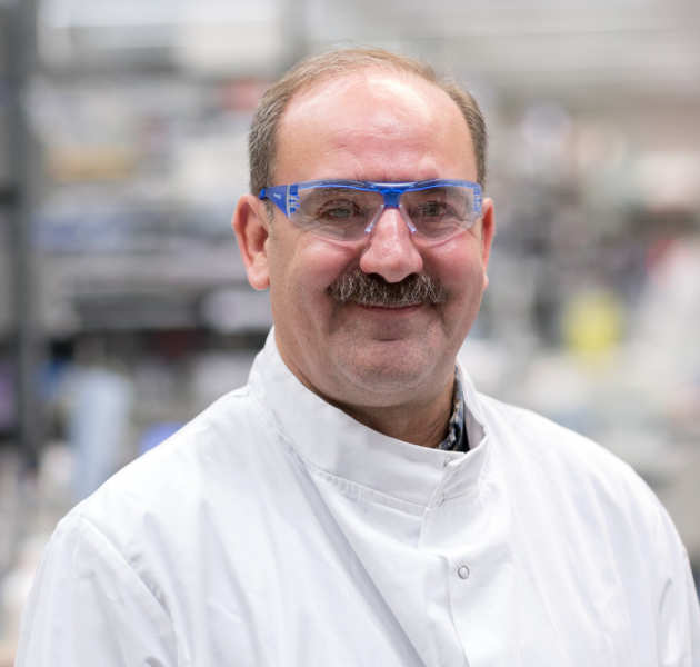
- Professor Harry Low
- Professor Paul Freemont
- Professor Xiaodong Zhang
- Professor Dale Wigley
- Dr David Riglar
- Dr Szymon Manka
- Dr Ravinash Krishna Kumar
- Dr Srinivasan Rengachari
 Professor Harry Low’s research team aims to understand how bacteria cause human disease and to develop novel antibiotics.
Some bacteria cause infections in humans that kill millions annually. To trigger the infection, the bacteria contain nanomachines within their cell surface that act like miniature pumps.These are secretion systems. One role of secretion systems is to deliver harmful proteins from inside the bacteria to external environments such as your cells. These harmful proteins act as molecular weaponry in many ways. They can form grappling hooks to help the bacteria attach to your internal surfaces or as molecular malware that maliciously reprogram your cells. We use powerful techniques such as cryo-electron microscopy and X-ray crystallography to visualise the precise position of the atoms within the secretion systems. In this way we can determine their 3D structure and chemistry. By understanding how the secretion systems work we have a much greater chance of developing medicines that stop the secretion systems from triggering human illness.

Professor Freemont's research focuses on two main areas – (1) understanding the molecular mechanisms of a number of protein complexes associated with cancer and bacterial infection using the tools of integrated structural biology and (2) development of synthetic biology tools and approaches for biosynthetic pathway engineering and biosensor development. His current interdisciplinary and collaborative research program focuses on understanding the molecular mechanisms of p97 and its role in mammalian proteostasis, the type VI secretion system from the human pathogen Pseudomonas aeruginosa and a number of bacterial signalling systems. His synthetic biology research is focused on cell-free systems for genetic circuit design and prototyping as well as also low-cost biosensor developments for healthcare applications. His lab is also engineering a number of biosynthetic pathways to produce complex molecules for potential therapeutic applications.
 Professor Zhang's research programme focuses on unravelling the mechanisms of macromolecular machines using a range of structural biology techniques including X-ray crystallography and cryo-electron microscopy. She is particularly interested in large macromolecular assemblies involved in DNA processing, as well as the AAA (ATPase Associated with diverse cellular Activities) protein family.
At present, her research focuses on three main areas: key components in DNA damage response, transcriptional regulation in bacteria, structure and mechanism of multifunctional p97
 Professor Dale Wigley and his team are finding out what happens when the DNA in a cell is damaged, and how the cell deals with it. If DNA damage is not repaired correctly, mistakes occur in important genes which can lead to cancer.
Professor Wigley studies bacteria grown in the lab to investigate DNA repair. Bacteria are a simple model system and easier to work with than human cells. Understanding the DNA repair processes in bacteria will help Professor Wigley and his team gain insight into the similar, more complex, processes at work in human cells.
Learning more about how cells repair damage to their DNA could lead to new ways to prevent or treat cancer in the future.
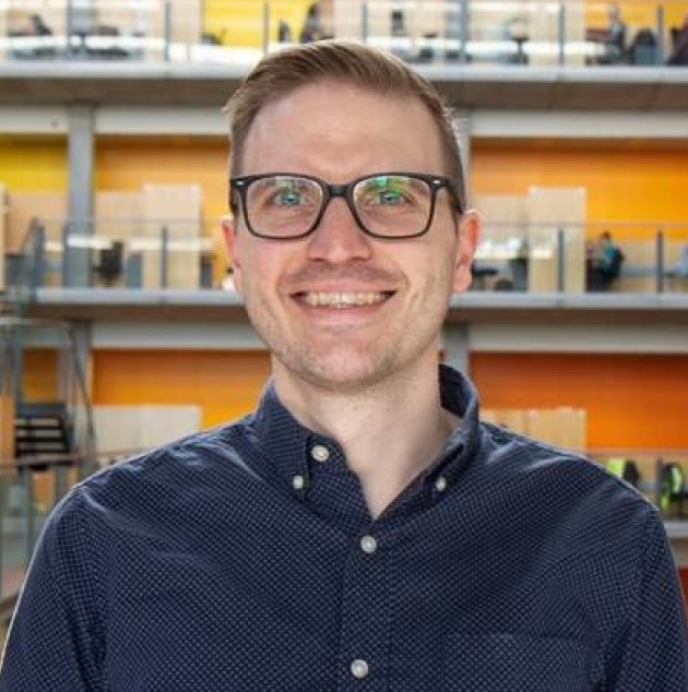 Dr David Riglar’s research aims to better understand the function of the gut and its resident microbiota during health and disease using a combination of synthetic biology, imaging and sequencing-based approaches. In particular, David is interested in how bacteria function differently in different regions of the gut.
The microbes that live in our guts are essential for maintaining our health. Changes to the make-up and function of the microbiota have been associated with a wide range of diseases including inflammatory bowel disease, colorectal cancer, diabetes and neurodevelopmental disorders.
Because of the intimate relationship they have with their surroundings, our microbes can be used to gain new insights into the gut and could one day diagnose, monitor and treat disease in the clinic. To this end, David’s lab is developing innovative technologies using engineered bacteria to probe and control the gut environment.
Dr. Manka's research focuses on elucidating the structural mechanisms underlying prion pathogenicity and evolution, with broader relevance to amyloid-driven diseases. His lab combines cryo-correlative light and electron microscopy (cryo-CLEM), cryo-electron tomography (cryo-ET), and single-molecule imaging to study protein aggregation at high resolution in situ.
By integrating genetic code expansion and bioorthogonal labelling, the lab is advancing the synthetic and structural cell biology toolkit to probe prion biology in unprecedented detail. The Manka Lab, supported by the UK Medical Research Council and the CJD Foundation, contributes to a deeper understanding of neurodegenerative disorders and systemic amyloidoses.
Find out more on the Manka Lab website

General enquiries
Section Manager
Brett Onslow
+44 (0)20 7594 3871
Personal Assistant for the Section of Structural Biology
Kasia Pearce
+44 (0)20 7594 2917
Laboratory Manager
Soo Mei Chee