Imperial researchers develop new ways to image atoms at ultra-low temperatures
/prod01/channel_4/imperial-news/media/news-images/3000X2000-main-article-images/3000-X-2000-News-article-images-7.jpg)
PhD student Mariana Palos in Imperial’s Department of Materials has developed new approaches to atomic-resolution electron microscopy at very low temperatures, allowing researchers to study materials as new physical properties emerge.
It was surprising to see how much more we could understand about a material by changing something as simple as the direction in which we scan it. Mariana Palos Research Postgraduate student in the Department of Materials
Mariana first joined Imperial as an MSc student through the British Council Women in STEM Scholarship programme, before staying on for a PhD in Dr Shelly Conroy's group in the Department of Materials, funded by the Royal Society.
Her first paper explored how changing the way we traditionally scan a sample on an electron microscope could address two persistent challenges in electron microscopy: sample movement during atomic-resolution imaging and damage caused by the electron beam. Both challenges become particularly important at very lowtemperatures, where maintaining a stable sample and controlling electron-beam exposure can be especially difficult.
Mariana programmed the microscope to scan in different ways, including spiral patterns, and found that the direction of the scan could help improve stability and reduce beam damage.
“It was surprising to see how much more we could understand about a material by changing something as simple as the direction in which we scan it. We found that different scan patterns can help with both stability and beam damage, which could open up new possibilities for studying beam-sensitive materials, from more complex materials to biological samples,” said Mariana.
Taking atomic-resolution microscopy to even colder temperatures
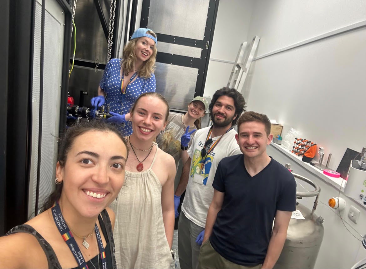
While finishing that work at liquid-nitrogen temperatures, Mariana was already working with Dr Noah Schnitzer on the next challenge: could we do ptychography at liquid-helium temperatures?
Mariana and Noah became joint first authors on the first demonstration in the world of atomic-resolution electron ptychography at around 20 K.
We can now start looking at materials atom by atom in phases that simply aren't there at room temperature. Mariana went from figuring out how to make these experiments more stable at liquid nitrogen to establish the first ptychography at helium temperatures. Dr Shelly Conroy Associate Professor in the Department of Materials
The work, supported by Dr Conroy's ERC Consolidator Grant, is important because many of the properties we care about in quantum materials only appear when they are very cold. If we want to understand why these materials behave as they do, we need to be able to see their atoms at the temperatures where the interesting physics is actually happening.
“This is the bit I find really exciting,” said Dr Conroy. “We can now start looking at materials atom by atom in phases that simply aren't there at room temperature. Mariana went from figuring out how to make these experiments more stable at liquid nitrogen to establish the first ptychography at helium temperatures.”
The advances in helium-temperature electron microscopy, including the work from Conroy's group at Imperial, were recently highlighted by Nature (https://www.nature.com/articles/d41586-026-02942-2 ).
Together, these advances are helping establish the UK as a leader in low-temperature, atomic-resolution characterisation, building the microscopy capabilities needed to understand and develop materials for the growing quantum technology sector.
Article text (excluding photos or graphics) © Imperial College London.
Photos and graphics subject to third party copyright used with permission or © Imperial College London.
Article people, mentions and related links
Sanjana Kakar
Faculty of Engineering
