BibTex format
@article{Chen:2010,
author = {Chen, S and McDowall, A and Dobro, MJ and Briegel, A and Ladinsky, M and Shi, J and Tocheva, EI and Beeby, M and Pilhofer, M and Ding, HJ and Li, Z and Gan, L and Morris, DM and Jensen, GJ},
journal = {Jove-Journal of Visualized Experiments},
title = {Electron cryotomography of bacterial cells},
url = {http://hdl.handle.net/10044/1/31159},
volume = {39},
year = {2010}
}
RIS format (EndNote, RefMan)
TY - JOUR
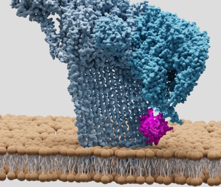
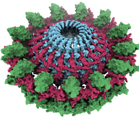
AB - While much is already known about the basic metabolism of bacterial cells, many fundamental questions are still surprisingly unanswered,including for instance how they generate and maintain specific cell shapes, establish polarity, segregate their genomes, and divide. In orderto understand these phenomena, imaging technologies are needed that bridge the resolution gap between fluorescence light microscopy andhigher-resolution methods such as X-ray crystallography and NMR spectroscopy.Electron cryotomography (ECT) is an emerging technology that does just this, allowing the ultrastructure of cells to be visualized in a near-nativestate, in three dimensions (3D), with "macromolecular" resolution (~4nm).1, 2 In ECT, cells are imaged in a vitreous, "frozen-hydrated" state ina cryo transmission electron microscope (cryoTEM) at low temperature (< -180°C). For slender cells (up to ~500 nm in thickness3), intact cellsare plunge-frozen within media across EM grids in cryogens such as ethane or ethane/propane mixtures. Thicker cells and biofilms can alsobe imaged in a vitreous state by first "high-pressure freezing" and then, "cryo-sectioning" them. A series of two-dimensional projection imagesare then collected through the sample as it is incrementally tilted along one or two axes. A three-dimensional reconstruction, or "tomogram" canthen be calculated from the images. While ECT requires expensive instrumentation, in recent years, it has been used in a few labs to reveal thestructures of various external appendages, the structures of different cell envelopes, the positions and structures of cytoskeletal filaments, andthe locations and architectures of large macromolecular assemblies such as flagellar motors, internal compartments and chemoreceptor arrays.1,2In this video article we illustrate how to image cells with ECT, including the processes of sample preparation, data collection, tomogramreconstruction, and interpre
AU - Chen,S
AU - McDowall,A
AU - Dobro,MJ
AU - Briegel,A
AU - Ladinsky,M
AU - Shi,J
AU - Tocheva,EI
AU - Beeby,M
AU - Pilhofer,M
AU - Ding,HJ
AU - Li,Z
AU - Gan,L
AU - Morris,DM
AU - Jensen,GJ
PY - 2010///
SN - 1940-087X
TI - Electron cryotomography of bacterial cells
T2 - Jove-Journal of Visualized Experiments
UR - http://hdl.handle.net/10044/1/31159
VL - 39
ER -