"Nanobiopsy" allows scientists to operate on living cells
by Sam Wong
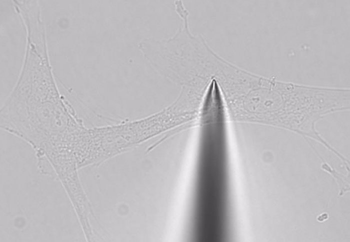
The technique uses a nanopipette to suck out minute volumes of a cell's contents.
Scientists have developed a device that can take a "biopsy" of a living cell, sampling minute volumes of its contents without killing it.
Much research on molecular biology is carried out on populations of cells, giving an average result that ignores the fact that every cell is different. Techniques for studying single cells usually destroy them, making it impossible to look at changes over time.
The new tool, called a nanobiopsy, uses a robotic glass nanopipette to pierce the cell membrane and extract a volume of around 50 femtolitres – 0.00000000000005 litres, around one per cent of the cell’s contents.
It will allow scientists to take samples repeatedly, to study the progression of disease at a molecular level in an individual cell. It can also be used to deliver material into cells, opening up ways to reprogram diseased cells.
“This is like doing surgery on individual cells,” said Dr Paolo Actis, from the Department of Medicine at Imperial College London, who developed the technology with colleagues at the University of California, Santa Cruz.
“This technology will be extremely useful for research in many areas. You could use it to dynamically study how cancer cells are different from healthy cells, or look at how brain cells are affected by Alzheimer’s disease. The possibilities are immense.”
To get inside the cell, the nanopipette is plunged downwards about one micrometre to pierce the cell membrane. Applying a voltage across the tip makes fluid flow into the pipette. When the pipette is removed from the cell, the membrane remains intact and the cell retains its shape.

The device is based on a scanning ion conductance microscope, which uses a robotic nanopipette, about 100 nanometres in diameter, to scan the surface of cells. The nanopipette is filled with an electrolyte solution and the ion current is measured inside the tip. When the pipette gets close to a cell membrane, the ion current decreases. This measurement is used to guide the tip across the surface of a sample at a constant distance, producing a picture of the surface.
In an initial study published in the journal ACS Nano, the researchers used the nanobiopsy technique to extract and sequence messenger RNA, molecules carrying genetic code transcribed from DNA in the cell’s nucleus. This allowed them to see which genes were being expressed in the cell.
They were also able to extract whole mitochondria – the power units of the cell. Mitochondria contain their own DNA, and the researchers discovered that the genomes of different mitochondria in the same cell are different.
They are now working on adapting the technology to incorporate sensors on the pipette tip that can instantly measure different molecules.
The research was funded by the US National Cancer Institute and National Institutes of Health.
Reference
P. Actis et al. ‘Compartmental Genomics in Living Cells Revealed by Single-Cell Nanobiopsy.’ ACS Nano, 2013. DOI: 10.1021/nn405097u
Article text (excluding photos or graphics) © Imperial College London.
Photos and graphics subject to third party copyright used with permission or © Imperial College London.
Reporter
Sam Wong
School of Professional Development