Miniature Fibre-Optic Probes for Rapid Bacteria Detection
The Hamlyn Centre researchers have realised a simple new technique to fabricate fibre‐optic probes, aiming to aid the diagnosis of lung infections.
The probes use a technique called Raman spectroscopy and are thin enough to be inserted into the body through endoscopes, catheters or needles.
Raman spectroscopy is a powerful analytical tool that has been adopted for identification and characterisation of materials in a wide range of applications. In the field of medicine, it has been spotlighted for detection and diagnosis of diseases ranging from infection to cancer. Using Raman spectroscopy to provide diagnostics in real-time and in a minimally invasive format would have many potential clinical benefits and this can be achieved by integrating miniature fibre-optic Raman probes into existing endoscopes.
However, deploying Raman spectroscopy in the clinic is challenging. Raman signals are usually weak and can easily be swamped by interfering background signals. To combat this problem, metal surfaces with micro- and nanoscale structures can provide a significant signal enhancement effect known as “surface-enhanced Raman spectroscopy (SERS).” Although this technique is well established, developing miniature fibre-optic SERS probes can be difficult due to the need for advanced micro-fabrication technologies.
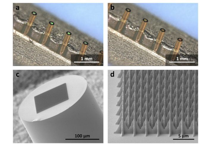
Researchers in the Hamlyn Centre have developed a new protocol for simple, effective and reliable fabrication of miniature fibre-optic SERS probes. This approach is based on a microscale 3D-printing technique known as two-photon polymerisation (2PP), which is commercially available (Nanoscribe GmbH, Germany). Using this approach, polymer microstructures are 3D-printed on the tip of an optical fibre and are then coated with a thin layer of gold (50 nm) to produce the metal surfaces for Raman signal enhancement. The research team demonstrated that the technique is reliable and reproducible, and the fibres used are only 220 µm in diameter—roughly 2-3 times the thickness of a single strand of hair.
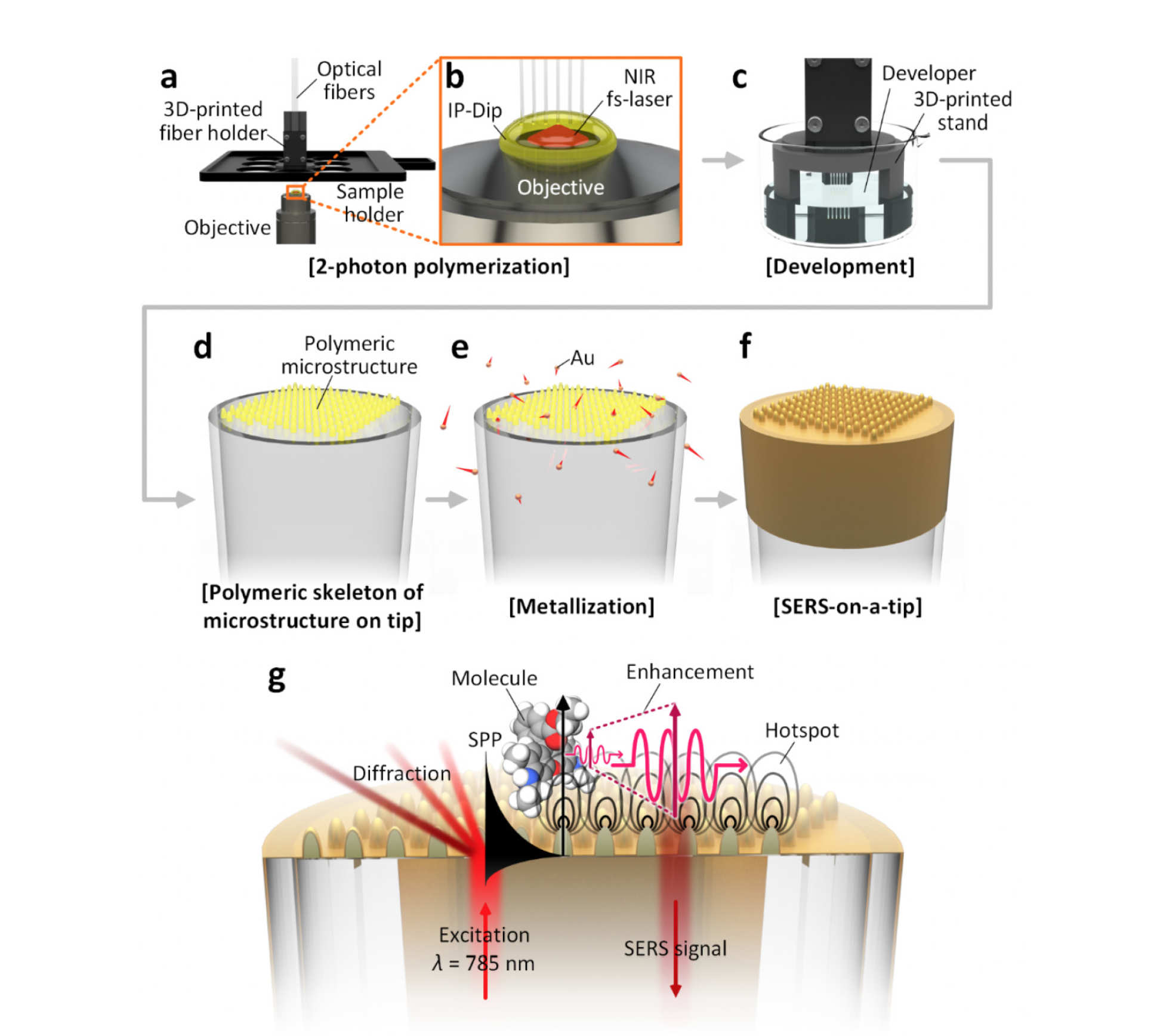
Having optimised the microstructures and characterised the sensing performance of the probes, the team also demonstrated the capability for rapid detection of bacteria in solution. This is the first report of detection of live, unlabelled bacteria using a fibre-optic SERS probe, and indicates the future potential for clinical applications.
The researchers are now continuing to further improve the sensing performance and robustness of their probes, with the hope that this will allow clinical testing—for example, for diagnosis of lung infections—in the future.
This research was conducted as a part of EPSRC Programme Grant, “Micro-robotics for Surgery (EP/P012779/1)” and was published in the early view of Advanced Optical Materials this week (J. Kim, D. J. Wales, A. J. Thompson, G.-Z. Yang, “Fiber-Optic SERS Probes Fabricated Using Two-Photon Polymerization For Rapid Detection of Bacteria”, Advanced Optical Materials 2020, 1901934).
Article supporters
Article text (excluding photos or graphics) © Imperial College London.
Photos and graphics subject to third party copyright used with permission or © Imperial College London.
Reporter
Dr Alex Thompson
Department of Surgery & Cancer
Dr Jang Ah Kim
Department of Mechanical Engineering
Erh-Ya (Asa) Tsui
Enterprise