HIV particles assemble ten times faster than previously estimated
Imperial researchers have developed a correlative life imaging technique that visualises real-time morphological changes when virus particles form.
Understanding the molecular mechanisms involved in the assembly of viruses is essential for discerning how viruses transmit from cell to cell and host to host. However, our current knowledge on the speed that individual virus particles assemble comes almost exclusively from life imaging – fluorescence microscopy. This technique requires fluorescent labelling of samples, which can affect virus particle assembly and release, causing inaccuracies in research.
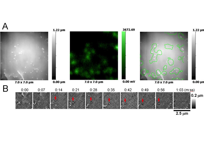
To better understand the molecular mechanisms involved in the assembly of viruses, researchers at Imperial College London, have pioneered an alternative, label-free, life imaging technique called Scanning Ion Conductance Microscopy (SICM) that enables them to visualise and follow the morphology of living cells with the resolution of Scanning Electron Microscope.
Using their novel SICM technique, the researchers followed the assembly of individual HIV particles on surfaces of living T-cells and measured the time required for their formation. Their results showed that on average the assembly of particles appeared to be ten times faster than when measured using previous techniques.
Importantly, SICM can also measure biophysical properties of cells such as stiffness, and be combined with fluorescence microscopy making it the only simultaneous correlative life imaging technique at present.
Speaking about the research, lead author from the Department of Metabolism, Digestion and Reproduction, Dr. Andrew Shevchuk said: "This new research tool enabled us to demonstrate that virus particle assembly can vary on different cell membrane domains and that tagging virus proteins with fluorescent labels e.g. green fluorescence protein (GFP) affects virus particle formation."
Although virus particle assembly is the very final stage of the entire virus replication cycle, and takes a fraction of the total time required, this research in fundamental bioscience highlights the importance of new research tools.
'Rapid formation of human immunodeficiency virus-like particles' by , , DOI: 10.1073/pnas.2008156117
Article text (excluding photos or graphics) © Imperial College London.
Photos and graphics subject to third party copyright used with permission or © Imperial College London.
Reporter
Benjie Coleman
Department of Surgery & Cancer