A Real-Time DRS Probe Tracking System for Gastrointestinal Cancer Detection
Researchers developed a probe tracking system for gastrointestinal cancer tissue detection using diffuse reflectance spectroscopy (DRS).
Gastrointestinal (GI) cancer remains a major contributor to the global cancer risk. When a surgery is carried out, the aim is to completely resect tumour tissues with clear margins, while preserving as much surrounding healthy tissues as possible. Patients with a positive circumferential resection margin (CRM) are reported to have poorer long-term survival and are more likely to have local recurrence of the tumour. It is therefore of prime importance to establish tissue margins accurately.
Diffuse reflectance spectroscopy (DRS) is a technique that allows discrimination of normal and abnormal tissue based on spectral data and has potential for cancer diagnosis. Nevertheless, "one major challenge of using DRS technique is that it is difficult for surgeons to build up a mental picture of the optical biopsy diagnosis when scanning across a large specimen, which makes it difficult for them to determine the resection margin", Professor Daniel Elson explained.
A Real-Time DRS Probe Tracking System for Gastrointestinal Cancer Tissue Detection
To overcome this limitation, the research team from the Hamlyn Centre and Department of Surgery and Cancer proposed a real-time DRS probe tracking system for GI cancer tissue detection and classification.
Lead researcher Ioannis Gkouzionis said: "The main purpose of this research is to use this proposed DRS probe tracking system to differentiate normal and tumour tissues in upper GI tissues in real-time, aiming to aid margin assessment during GI cancer surgery."
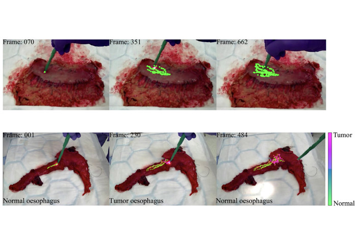
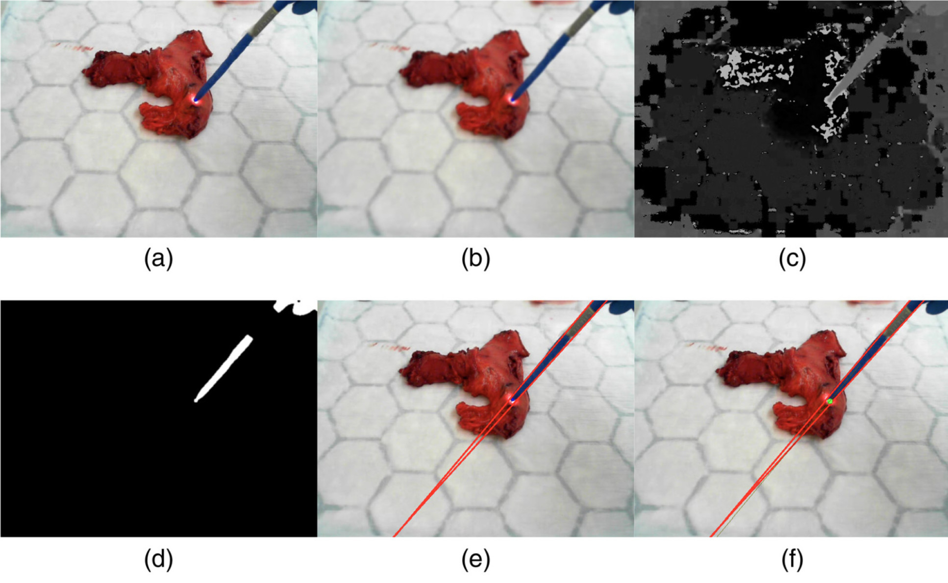
DRS fibre probe tracking procedure and detection workflow
The DRS set-up was used in the operating theatre during the gastrointestinal cancer procedures.
To validate the proposed tracking system, the team recruited 26 patients who were having undergoing gastric or oesophageal cancer resection surgery.
 After the specimen was resected, the team used the DRS probe to sample an area of normal tissue and the area of suspected tumour tissue. The tracking system was based on a green-coloured marker (which was wrapped around the distal end of the DRS probe).
Ex-Vivo Experimental Evaluation
The spectral data was captured on an easy-to-use graphical user interface in real-time and further analysed the data by using a machine learning classifier (Extreme Gradient Boosting) based on histopathology validation.
Ioannis Gkouzionis said: "Real-time tracking, combined with binary classification of normal or tumour tissue, allowed ones to have a clear on-screen visualisation of the tissue type, which can be used intra-operatively to aid margin assessment in real-time clinical practice."
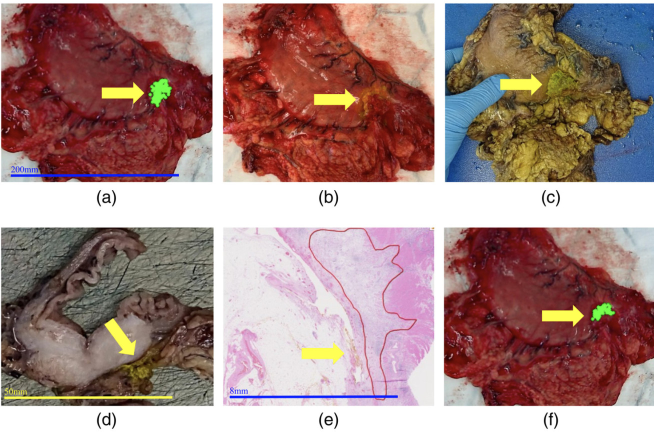
In conclusion, the proposed real-time DRS tracking system was validated on ex-vivo tissue with histological ground truth. An overall diagnostic accuracy of 94% for stomach cancer and 96% for the oesophageal cancer demonstrated the strength and clinical value of the technique.
"It is a fundamental step towards the development of a real-time in-vivo tumour mapping tool for oesophageal and gastric cancers to improve long-term outcomes", Clinical Research Fellow Scarlet Nazarian emphasised.
This research was supported by the National Institute for Health Research (NIHR) Imperial Biomedical Research Centre (BRC) and the Cancer Research UK (CRUK) Imperial Centre. (Ioannis Gkouzionis, Scarlet Nazarian, Michal Kawka, Ara Darzi, Nisha Patel, Christopher J. Peters, and Daniel Elson, "Real-time tracking of a diffuse reflectance spectroscopy probe used to aid histological validation of margin assessment in upper gastrointestinal cancer resection surgery", Journal of Biomedical Optics, 27(2), 025001, Feb 2022)
Article supporters
Article text (excluding photos or graphics) © Imperial College London.
Photos and graphics subject to third party copyright used with permission or © Imperial College London.
Reporter
Erh-Ya (Asa) Tsui
Enterprise