Single brain scan can diagnose Alzheimer’s disease
by Maxine Myers
A single MRI scan of the brain could be enough to diagnose Alzheimer’s disease, according to new research by Imperial College London.
The research uses machine learning technology to look at structural features within the brain, including in regions not previously associated with Alzheimer’s. The advantage of the technique is its simplicity and the fact that it can identify the disease at an early stage when it can be very difficult to diagnose.
Although there is no cure for Alzheimer’s disease, getting a diagnosis quickly at an early stage helps patients. It allows them to access help and support, get treatment to manage their symptoms and plan for the future. Being able to accurately identify patients at an early stage of the disease will also help researchers to understand the brain changes that trigger the disease, and support development and trials of new treatments.
Common form of dementia
The research is published in the Nature Portfolio Journal, Communications Medicine, and funded through the National Institute for Health and Care Research (NIHR) Imperial Biomedical Research Centre and the Medical Research Council.
Alzheimer’s disease is the most common form of dementia, affecting over half a million people in the UK. Although most people with Alzheimer’s disease develop it after the age of 65, people under this age can develop it too. The most frequent symptoms of dementia are memory loss and difficulties with thinking, problem solving and language.
Doctors currently use a raft of tests to diagnose Alzheimer’s disease, including memory and cognitive tests and brain scans. The scans are used to check for protein deposits in the brain and shrinkage of the hippocampus, the area of the brain linked to memory. All of these tests can take several weeks, both to arrange and to process.
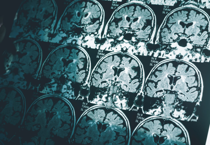
The new approach requires just one of these – a magnetic resonance imaging (MRI) brain scan taken on a standard 1.5 Tesla machine, which is commonly found in most hospitals.
The researchers adapted an algorithm developed for use in classifying cancer tumours, and applied it to the brain. They divided the brain into 115 regions and allocated 660 different features, such as size, shape and texture, to assess each region. They then trained the algorithm to identify where changes to these features could accurately predict the existence of Alzheimer’s disease.
Using data from the Alzheimer’s Disease Neuroimaging Initiative, the team tested their approach on brain scans from over 400 patients with early and later stage Alzheimer’s, healthy controls and patients with other neurological conditions, including frontotemporal dementia and Parkinson’s disease. They also tested it with data from over 80 patients undergoing diagnostic tests for Alzheimer’s at Imperial College Healthcare NHS Trust.
Single test
They found that in 98 per cent of cases, the MRI-based machine learning system alone could accurately predict whether the patient had Alzheimer’s disease or not. It was also able to distinguish between early and late-stage Alzheimer’s with fairly high accuracy, in 79 per cent of patients.
Professor Eric Aboagye, from Imperial’s Department of Surgery and Cancer, who led the research, said: “Currently no other simple and widely available methods can predict Alzheimer’s disease with this level of accuracy, so our research is an important step forward. Many patients who present with Alzheimer’s at memory clinics do also have other neurological conditions, but even within this group our system could pick out those patients who had Alzheimer’s from those who did not.
“Waiting for a diagnosis can be a horrible experience for patients and their families. If we could cut down the amount of time they have to wait, make diagnosis a simpler process, and reduce some of the uncertainty, that would help a great deal. Our new approach could also identify early-stage patients for clinical trials of new drug treatments or lifestyle changes, which is currently very hard to do.”
The new system spotted changes in areas of the brain not previously associated with Alzheimer’s disease, including the cerebellum (the part of the brain that coordinates and regulates physical activity) and the ventral diencephalon (linked to the senses, sight and hearing). This opens up potential new avenues for research into these areas and their links to Alzheimer’s disease.
Dr Paresh Malhotra, who is a consultant neurologist at Imperial College Healthcare NHS Trust and a researcher in Imperial’s Department of Brain Sciences, said: “Although neuroradiologists already interpret MRI scans to help diagnose Alzheimer’s, there are likely to be features of the scans that aren’t visible, even to specialists. Using an algorithm able to select texture and subtle structural features in the brain that are affected by Alzheimer’s could really enhance the information we can gain from standard imaging techniques.”
Article supporters
Article text (excluding photos or graphics) © Imperial College London.
Photos and graphics subject to third party copyright used with permission or © Imperial College London.
Reporter
Maxine Myers
Communications Division