Researchers trial tiny new microscope to detect breast cancer
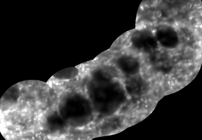
Microscope imaging of invasive breast cancer cells
An Imperial-developed tiny microscope that can be steered through small bodily spaces during surgery has entered its next phase of laboratory trials.
The endo-microscope – a microscope designed to be inserted into the body to provide views of tissue and organs – produces images with unprecedented speed. The researchers say the new technology, which is now undergoing laboratory testing on human cancer tissues, could potentially improve the diagnosis and treatment of breast cancer.
The multi-wavelength endo-microscope and accompanying image acquisition and classification software was developed as part of the five-year Micro-Robotics for Surgery programme by Dr Khushi Vyas and colleagues at Imperial College London. It is supported by the Engineering and Physical Sciences Research Council (EPSRC), part of UK Research and Innovation.
Using safe and rapid staining dyes, the endo-microscope could produce real-time histology-like images of tissue micro-architecture during surgery without the need to excise any tissue. If proved successful in human trials, it could reduce reoperation rates, unnecessary removal of healthy tissue and potentially increase how many patients can be seen.
The researchers have used their system for preliminary studies on human cancer tissue and are now testing its use by surgeons and pathologists on laboratory samples of cancerous tissue.
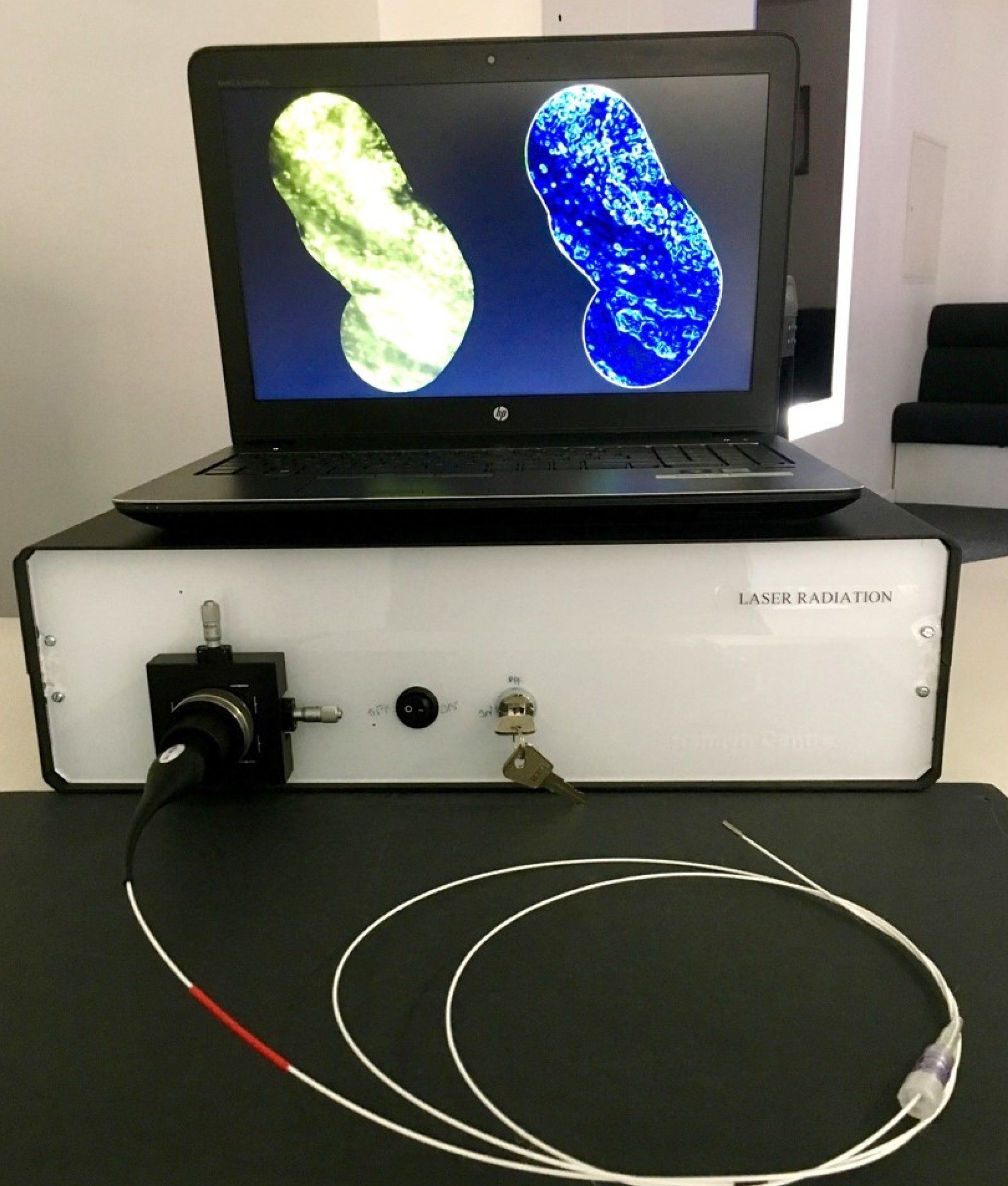
Dr Vyas of Imperial’s Department of Electrical and Electronic Engineering and Hamlyn Centre said: “A key focus of this EPSRC-funded work has been the development of hardware and accompanying tissue staining and classification protocols enabling the new system to generate diagnostic relevant tissue images at 120 frames per second – a huge leap forward in terms of image acquisition.”
Exploring small spaces
When exploring spaces such as breast ducts in preliminary studies on cancer tissue, the instrument was able to pinpoint features smaller than a single cell. In live surgery, it could aid high-precision breast-conserving surgery by enabling surgeons to identify, extremely accurately and much more quickly than currently possible, suspicious tissue around tumours as well as cancerous cells just a hundredth of a millimetre across. It could also be used in the lungs, urinary tract, digestive system and brain in the future.
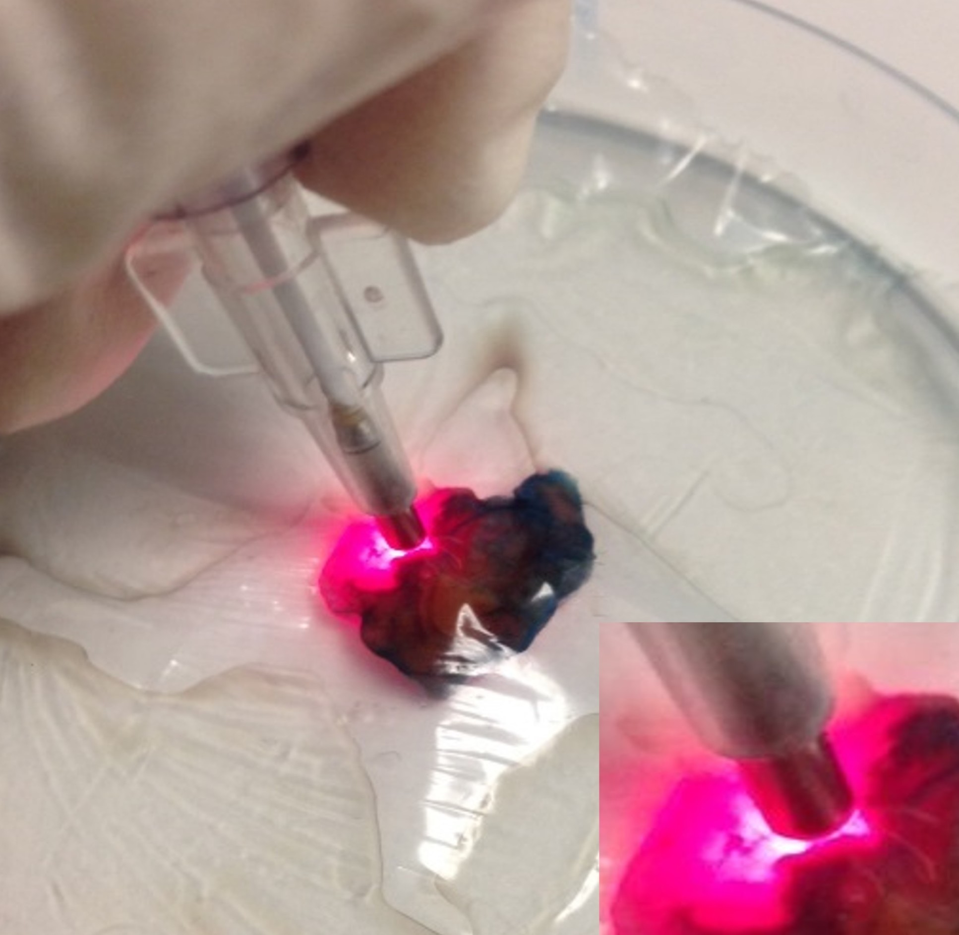
Compact, portable and easy to use, the endo-microscope comprises a tiny lens assembly fitted to the end of a flexible polymer fibre-bundle the width of 25-30 human hairs. The system is designed to be set up next to the patient in the operating theatre. The surgeon would carefully insert the fibre into the patient by hand, holding it like a pen; alternatively, the endo-microscope could be fitted into a robotic scanner to precisely scan the entire breast cavity for suspicious tissue.
The instrument can be easily steered through tissue, with instant large-area image generation of whatever the fibre-tip comes into contact with, similar to the panorama feature on smartphones. The high-resolution images are displayed in real time on a high-definition monitor that the surgeon consults as they work.
Principal Investigator on the project Professor Eric Yeatman, of Imperial’s Department of Electrical and Electronic Engineering, said: "This innovation resulted from the close collaboration of engineering, science and clinical researchers. We're very grateful to the EPSRC for making this collaboration possible through their Programme Grant funding."
EPSRC Director for Cross-Council Programmes, Dr Kedar Pandya, said: “By reducing the time it takes to identify cancerous cells and improve the accuracy of imaging the endo-microscope developed by Dr Vyas and her team could benefit patients and the NHS by reducing waiting lists.
“As we mark Breast Cancer Awareness Month this illustrates the important role that cutting-edge research and innovation will play in helping us to detect and treat the most common cancer in the UK.”
The new endo-microscope has been developed as part of the five-year EPSRC-funded Micro-Robotics for Surgery programme, led by Imperial College London and involving 12 partner organisations. Running from April 2017 to December 2022, the programme has received a total of £6.2 million in EPSRC support.
This story is adapted from a press release by EPSRC.
Images: Vyas et al.
Article supporters
Article text (excluding photos or graphics) © Imperial College London.
Photos and graphics subject to third party copyright used with permission or © Imperial College London.
Reporter
Caroline Brogan
Communications Division