Novel heart model creates lifelike 4D images for better cardiac research
by Gemma Ralton
Researchers from Imperial College London have created ‘CHeart’, a new machine learning model that generates 4D images of the human heart.
The new, state-of-the-art computer model offers insights into how clinical factors like age, gender and disease may influence the heart’s structure and function by generating realistic 4D images (segmentation maps) – something that currently is very difficult to study in real life due to a lack of extensive data.
“We have created a new tool for doctors and researchers to understand heart diseases better and to foresee how different clinical factors might affect the heart. This can lead to better diagnoses and personalized treatments, which could improve patient care significantly.” Dr Mengyun Qiao Lead Author
The team of researchers including the Data Science Institute’s Dr Mengyun Qiao and Dr Wenjia Bai used a dataset of 3D cardiac MR images acquired from Hammersmith Hospital, and cutting edge image segmentation methods to train the model to identify patterns in heart structure that deviate from the norm, helping to improve diagnostics and treatment for heart conditions.
According to Lead Author Dr Qiao: “We have created a new tool for doctors and researchers to understand heart diseases better and to foresee how different clinical factors might affect the heart. This can lead to better diagnoses and personalized treatments, which could improve patient care significantly.”
Their research is published in IEEE transactions on medical imaging.
Using computational tools for clinical applications
Cardiac imaging plays a crucial role in cardiovascular image diagnosis and management, and understanding the relationship between the 3D structure and movement of the heart and non-imaging clinical factors is important for improving our ability to diagnose and manage heart conditions using medical imaging.
Current approaches to studying the relationship between clinical factors like age, gender and disease on the heart’s structure and function are limited due to the lack of comprehensive and diverse cardiac imaging data.
It is a complex problem, but advanced computational tools can help to bridge spatial-temporal imaging features and non-imaging clinical factors.
The researchers proposed a conditional generative model as the solution – a type of machine learning model that can uses generative artificial intelligence technology to generate data samples based on specific conditions or inputs.
CHeart: A realistic computer model of the human heart
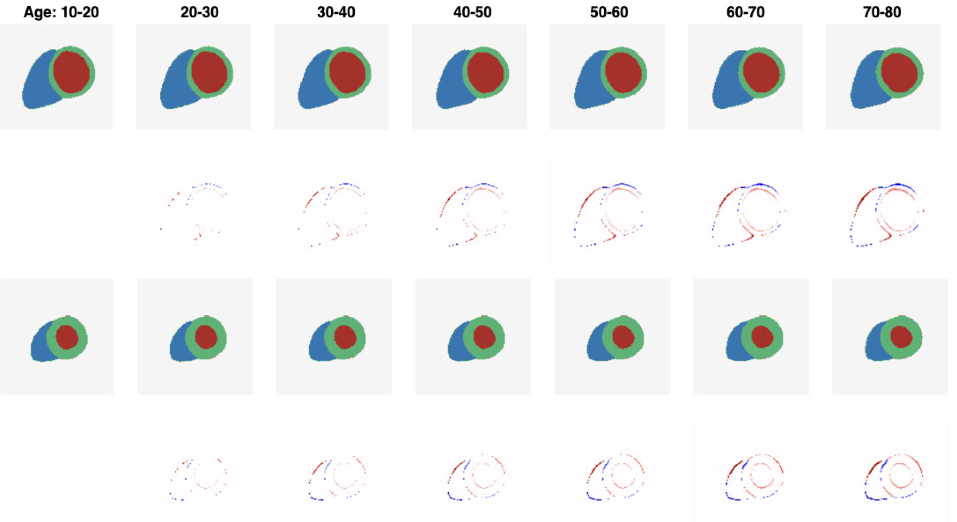
In the study, the researchers created a generative model ‘CHeart’ which produces realistic cardiac anatomical sequences based on non-imaging factors such as age, gender, weight, height and blood pressure.
In order to create CHeart, the researchers firstly gathered detailed 3D images of the heart from 1383 subjects using cardiac MRI. These images capture the heart at different times during its beating cycle, providing a dynamic sequence of how the heart looks and moves.
Using machine learning techniques, the team then fed the model with heart images and corresponding clinical data. Over time, the model ‘learned’ how a typical heart looks and how different factors might change its appearance.
According to Dr Qiao: “We validated our model by comparing the artificial heart images it produced with actual heart images, and the similarities were remarkably close, suggesting our model can be a reliable tool for studying the heart.”
Next steps
CHeart represents the first computer generative model to accurately generate 4D images of the human heart. However, the computational cost is currently quite high, due to the high dimensionality of the data.
Similarly, the study uses segmentation maps to represent cardiac anatomy, which simplifies the complex nature of cardiac structures to a degree that may omit relevant details. It helps to focus the model's learning on anatomical variations rather than image styles, but this representation might limit the model's capacity to capture every nuance of the cardiac anatomy.
Moving forward, Dr Qiao hopes to address these limitations by reducing the computational complexity and finding ways to enhance the anatomical representation within the model.
Regardless, CHeart demonstrates a unique application of generative models and their use in health sciences, offering a promising tool for clinicians and medical scientists to improve treatment for cardiac conditions.
-
‘CHeart: A Conditional Spatio-Temporal Generative Model for Cardiac Anatomy’ by Qiao et al., published on 10 November 2023 in IEEE transactions on medical imaging.
Header image sourced from Jesse Orrico, Unsplash.
Article text (excluding photos or graphics) © Imperial College London.
Photos and graphics subject to third party copyright used with permission or © Imperial College London.
Reporter
Gemma Ralton
Faculty of Engineering