Polarimetry is the study and measurement of the polarisation state of light and is a popular and useful tool in science today. Applications vary from astronomy, microscopy and biomedical diagnosis to more fundamental crystallographic, material and single molecule studies. Whilst the polarisation state of light itself, can be used to transmit information, hence presenting new possibilities in communications and optical data storage, changes in polarisation induced by an object can, alternatively, be used to expose new material or sample properties to scientific scrutiny, such material birefringence, chirality or molecular orientation. Our current polarimetry research includes the following:
1. Polarisation microscopy
Incorporating polarisation sensitive elements into existing optical imaging systems enables polarisation imaging, in which either the polarisation state of the incident light is mapped (Stokes imaging) or the polarisation properties of the object are considered (Mueller matrix imaging). High resolution imaging, however requires high numerical aperture lenses, which further transform the polarisation. Development of accurate theoretical models and experimental calibration procedures, is thus important for accurate quantitative polarisation measurements. Recent decades have also seen a number of single molecule imaging techniques, such as fluorescence microscopy, STED and other localisation imaging modalities. Such techniques typically only measure the optical intensity from a molecule and eschew the information carried by the polarisation of light. We, however, consider how polarisation measurements can be used to yield additional information, for example for determining the 3D orientation of single fluorescent molecules, important in, for instance, studies of cell dynamics and monitoring their local environmental conditions.
2. Optimisation of polarimeters
In biological applications light levels areoften kept relatively low so as to avoid sample damage, whereas in quantum or astronomically applications photon numbers are frequently inherently low. Consequently, the presence of noise, be it fundamental shot noise or technical noise from scattering, can be quite detrimental to measurement accuracy. In polarimetry, this problem is further compounded by the need to make multiple measurements of different polarisation states. Optimising the design of polarimeters is thus an important component of high accuracy studies. Using elements of discrete computational geometry, information and statistical estimation theory we consider how improvements can be made through use of a priori knowledge of the system, choice of different measurement bases and enforcing physical constraints.
3. Spectropolarimetry
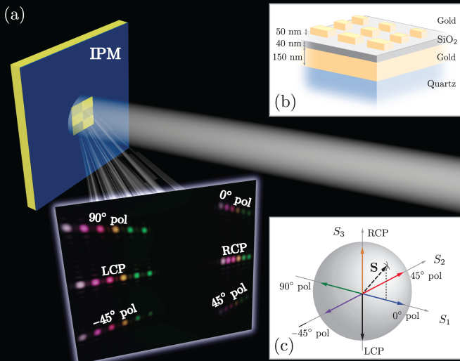
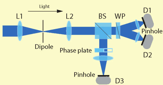
In addition to the additional information afforded by polarisation measurements, spectral degrees of freedom can yield details of the chemical composition of a sample, particle velocity, local magnetic and electric fields and more. Spectral-polarimetry therefore constitutes an exhaustive analytic optical tool. Spectropolarimetry can be practically realized using a number of experimental architectures, which differ through the means of spectral and polarization discrimination. Separation of individual spectral components is typically achieved by means of either a tunable optical or a dispersive element, such as a prism or grating structure. On the other hand, differing polarization components, are typically found using sequential measurements with variable polarisation elements or simultaneously using a division of amplitude polarimeter or polarization gratings. We have recently demonstrated a novel architecture employing plasmonic metasurfaces (nano-structured thin metallic films) enabling simultaneous determination of both spectral and polarisation information using a compact device, promising many applications in polarization imaging, quantum communication and quantitative sensing. Capitalising on our work on Müller matrix microscopy, we are also developing a spectroscopic version of this microscope that will permit 3D functional imaging of plasmonic structures and photonic crystals.