One of the main issues with Brillouin imaging is the coupling between the acoustic velocity and the refractive index within the sample when measuring the Brillouin frequency shift. This prevents obtaining quantitative data of the actual mechanical properties of the sample if the refractive index is unknown. Aiming to solve this problem, the combination of Brillouin and phase imaging systems is a promising assembly for separately monitoring the mechanical and optical properties of biological samples.
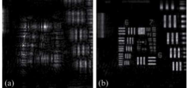 Fig. 1 Numerical refocusing of the amplitude of a test target USAF 1951 by means of digital holographic microscopy: (a) Reconstructed intensity in the camera plane. (b) Numerically refocused intensity distribution in the image plane.
Phase imaging techniques tackle the problem of measuring variations in the optical path length of light that passes through a given sample. When applied to microscopy and, especially, to biological specimens, these techniques are a unique tool for obtaining well-contrasted images of label-free samples. There are mainly two groups of phase-imaging techniques: the first group, known as phase-contrast techniques, involves the acquisition of qualitative information of phase changes in the sample, which is typically related with the derivative of the phase. The second group allows retrieving the quantitative information of the phase (by means of a single-shot in some cases) and are based on interferometric-holographic acquisition of the field scattered by the sample. By use of this information, the whole complex amplitude distribution of the field can be recovered, enabling both quantitative phase imaging and numerical refocusing of the field to be done. One of the most suitable techniques for performing this task is digital holographic microscopy, which only requires recording the interference between a beam diffracted by the sample and a free-space propagated beam. If the thickness of the sample is known, the phase-map can be transferred into an index of refraction map of the sample, which is the goal in terms of decoupling the index of refraction and the acoustic velocity when studying the Brillouin frequency shift.
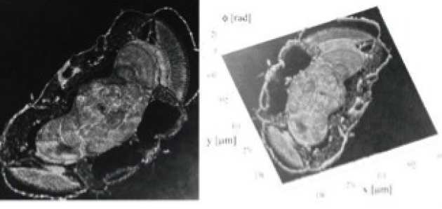 Fig. 2 Quantitative phase image of a drosophila melanogaster head cut obtained by digital holographic microscopy.
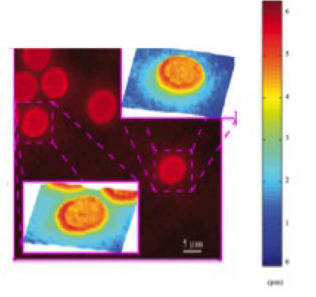
Fig. 3 Quantitative phase image of a red blood cell smear obtained by means of a digital holographic microscope.