By Christopher Uy
In a paper recently published in JACC Cardiovascular Imaging, we presented a series of images which summarise the various imaging modalities utilised in the management of Takayasu’s arteritis (TA)—a rare autoimmune vasculitis which affects the aorta and its major branches. Immunosuppressive therapy is employed to suppress inflammation, to prevent arterial injury and encourage positive remodelling. These images represent the 20-year cumulative experience of the multidisciplinary team at Imperial College NHS Trust treating this rare disease.
The natural history of TA is apparent through these images as we outline its diagnosis, extent and pattern, monitoring, and long-term outcomes. Early diagnosis can be facilitated with positron-emission tomography, magnetic resonance imaging (MRI) / angiography (MRA), computed tomography angiography, and high-resolution ultrasound. Furthermore, MRI and MRA remain invaluable for interval monitoring for treatment responses and promptly detecting signs of disease progression without ionising radiation. Other specialised imaging modalities are occasionally useful such as Tc99m-DSMA radionucleotide scanning to determine differential renal function in those with renal artery stenosis, and Tc99m HMPAO SPECT imaging to assess cerebral perfusion for those presenting with cerebral ischaemia, respectively. Integrated non-invasive imaging has transformed the approach to TA allowing for optimised clinical care in this patient cohort.
Link to publication: https://pubmed.ncbi.nlm.nih.gov/32682724/
References
1. Uy CP, Tarkin J, Gopalan D, Barwick T, Tombetti E, Youngstein T, Mason JC. The Impact of Integrated Noninvasive Imaging in the Management of Takayasu Arteritis. JACC Cardiovascular Imaging. 2020.
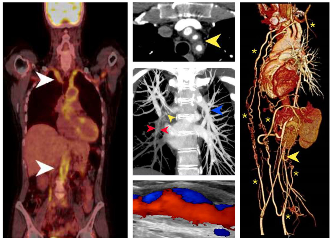
A. FDG-PET/CT imaging demonstrates intense homogenous tracer uptake throughout the vessel wall, consistent with aortitis and subclavian arteritis (arrows). B. Supra-aortic artery arteritis, with cuffing of the great vessels by inflammatory tissue is shown here by Computed tomography (CT) angiography. C. Pulmonary artery involvement is seen in up to 50% of TA patients. This CT pulmonary angiogram demonstrates severe stenosis of the right lower lobe pulmonary artery (red and yellow arrows). D. This high resolution ultrasound image with colour Doppler reveals concentric, homogenous thickening of the common carotid artery wall with evidence of focal arterial wall dilatation and disturbed blood flow. E. Tissue ischemia may be an important driver for collateral artery formation which can significantly reduce ischemic symptoms. The thoracic MRA 3D volume rendered reconstruction shows extensive disease of the aorta culminating in distal occlusion (arrow) and extensive collaterals (asterisks) which revascularise the gastrointestinal and the lower limb circulation.
General enquiries
For any questions related to the Centre, please contact:
Vasculitis Centre of Excellence Admin
VasculitisCoE@imperial.ac.uk
