Leica Cell DIVE - a robust approach to whole tissue imaging
- Built for Life Science Researchers: a user friendly yet highly stable system designed for studying tissues in detail.
- Whole tissue imaging: uses a widefield microscope - especially useful for understanding where cells are located and how they interact in tissue samples (spatial biology).
- Fully automated: works with the BioAssemblyBot®200 to automatically stain, image, and process multiple slides —even runs 24/7.
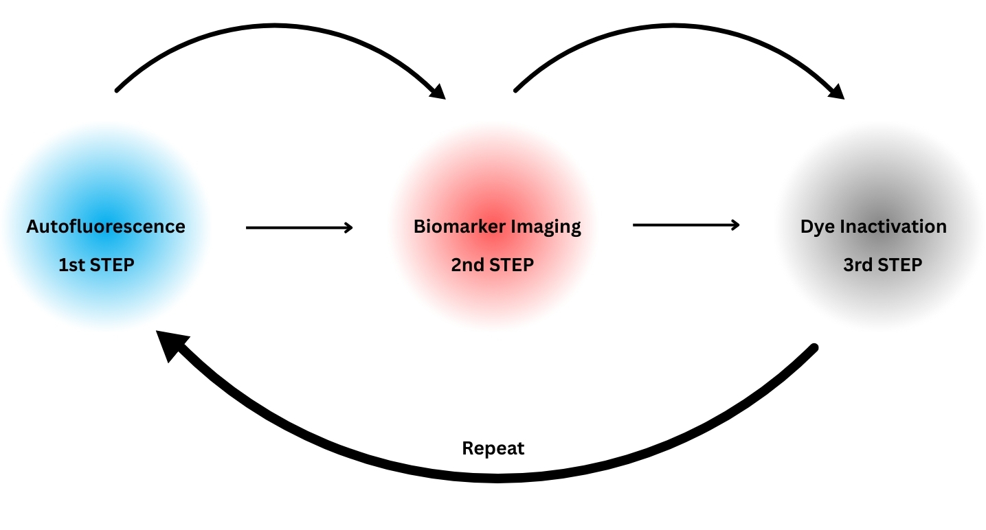
How it works
Traditional methods:
- All targets are labelled and imaged in one go
- Multiplexing can be difficult because the fluorescent dyes can overlap and interfere with each other.
- Requires technical expertise to separate similar dyes
Cell Dive’s approach:
- Labels and images 4 targets at a time, multiplexing is achieved via multiple rounds of labelling and imaging.
- This avoids overlap and makes it easier to study many different markers clearly.
- Simple concept thus no expertise required
Advantages of this microscope
- Easy to use: you don’t need to be a microscopy expert to get great results.
- Hands-free operation: the system focuses and runs on its own — no need for constant supervision.
- High capacity: can analyse over 60 different markers in a single tissue sample — helping researchers understand complex cell types and functions.
- Clear images: produces high-quality, full-tissue images that look like traditional H&E stains.
- Timesaving: the robot handles the work, so you don’t have to stand by the microscope.
- Reliable results: built-in calibration ensures consistent and accurate imaging every time.
Location
Room 304B, Imperial Centre for Translational and Experimental Medicine - ICTEM
Hammersmith Campus
Du Cane Road, London, W12 0NN
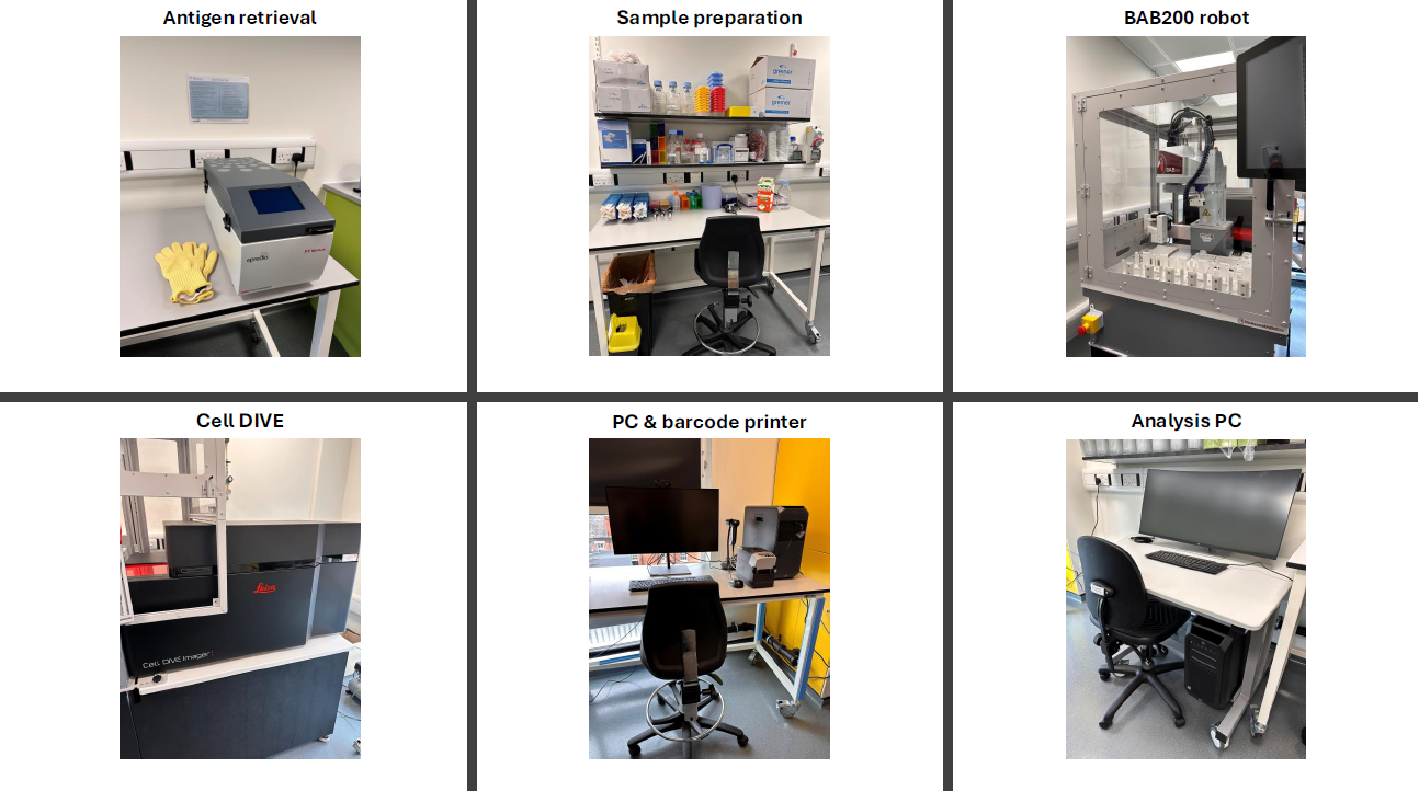
Facilities available in room 304B:
Quickstart guides
Quickstart guide - HMI1 - Leica Cell DIVE Multiplex Imager
Quick start guide - BioAssemblyBot 200 (BAB200)
Objectives for imaging
- 2x CFI Plan Apo Lambda (general overview)
- 10x CFI Plan Apo Lambda (region of interest selection)
- 20x CFI S Plan Fluor LWD (final imaging)
Light source and emission filter
| LEDs | Central Excitation Wavelength | Emission Filter |
|---|---|---|
| Blue | 390 | 411–446 (DAPI or similar) |
| Green | 470 | 495–512 (FITC or similar) |
| Orange | 542 | 574–599 (Cyanine 3 or similar) |
| Far red | 631 | 664–690 (Cyanine 5 or similar) |
| Near IR | 730 | 772–814 (Cyanine 7 or similar) |
Validated antibodies
Antibodies validated for use on the Cell DIVE.
Leica Microsystems, Cell DIVE - Antibodies in Multiplexed Imaging
Technical notes
Leica Microsystems Cell DIVE Technical Note
Leica Microsystems Cell DIVE Application Notes
Leica webinars
Transforming tissue research with open multiplexing
Automated Multiplexed Staining and Imaging with Cell DIVE
Offline software
Booking guidelines, Charges, make a booking
General enquiries
FILM
Sir Alexander Fleming Building
South Kensington Campus
Imperial College London
Exhibition Road
London SW7 2AZ, UK
film-service@imperial.ac.uk