Liver cancer cell ‘switch’ found that could improve future therapies
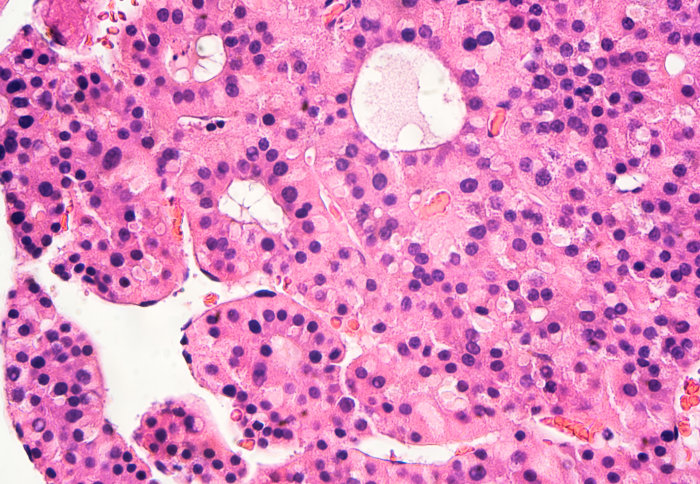
Liver cells with hepatocellular carcinoma (HCC)
Imperial researchers have found a new cell mechanism that could be used to target tumours in the future.
The newly discovered ‘switch’ controls the activity of two proteins – ‘deleted in liver cancer-1’ (DLC1), a tumour suppressor, and talin. Both proteins control whether or not cancer cells spread to other parts of the body, but the exact mechanisms have remained unclear.
Now, a team of researchers led by Dr Armando E del Río Hernández from Imperial’s Department of Bioengineering have found that the way talin unfolds in response to movement is the determining factor in DLC1 activity.
Our findings have implications for tissue engineering, cancer research, and drug testing using organ-on-chip technology. Dr Armando E del Río Hernández Department of Bioengineering
It is therefore crucial for the development of hepatocellular carcinoma (HCC) – the most common type of liver cancer.
Under the right conditions, such as certain gene activation or lowered immune system, cancer cells can migrate or ‘metastasise’ and invade other tissues, which spreads the cancer. Once cancer spreads to secondary sites in the body, it is much harder to treat – so preventing this is a priority for cancer researchers.
The scientists say their findings in this paper, published in PLOS Biology, help us understand how tumour cells migrate and therefore how to target and prevent cancer spread, particularly in HCC.
Dr Hernández said: “Our findings have implications for tissue engineering, cancer research, and drug testing using organ-on-chip technology.”
Protein performance
Like a skeleton controlled by outside mechanical forces, cells contract and move in response to outside forces. Cells’ inner structures, known as cytoskeletons, produce the forces that lead to cell migration in cancer cells.
The talin protein attaches to the cell membrane and creates a bridge between the outside and inside of the cell. It is coiled like a spring so that it can sense and respond to mechanical forces. These forces change the shape of talin, which deactivates DLC1 so that it cannot perform its tumour suppressing fuction, leading to cancer. Until now, researchers did not know exactly how talin changed shape.
Dr Hernández and colleagues from the University of Tampere, Finland, studied talin’s response to movement in fibroblasts – cells that help to heal wounds and repair damaged tissues. When unchecked, fibroblasts can produce scar tissue in the liver, which leads to cirrhosis and HCC.
Next, they will look into the broader implications of finding the ‘switch’. Co-lead author Dr Vesa Hytönen, from the University of Tampere said: “Although we looked only at fibroblasts, all molecules included in this study are found widely throughout the body. We’re therefore hopeful that this mechanism will be found in other tissues.”

The research was funded by the European Research Council and Academy of Finland.
“Mechanotransduction in talin through the interaction of the R8 domain with DLC1” by Haining AWM, Rahikainen R, Cortes E, Lachowski D, Rice A, von Essen M, et al. Published 20 July 2018 in PLOS Biology.
Image credit: Shutterstock/David Litman
Article supporters
Article text (excluding photos or graphics) © Imperial College London.
Photos and graphics subject to third party copyright used with permission or © Imperial College London.
Reporter
Caroline Brogan
Communications Division