New way to look at cell membranes could change the way we study disease
A new technique to study intact parts of cell membranes could revolutionise studies of cancer, metabolic and heart diseases.
Membranes protect all of our cells and the organelles inside them, including the mitochondria – the powerhouse of the cell. These membranes are studded with biological machinery made of proteins that enable molecular cargo to pass in and out.
With the development of this method, the application of mass spectrometry in biology will be taken to a new level, using it to make discoveries that would not have been possible before. Professor Steve Matthews
Studying these membrane-embedded machines in their native state is therefore crucial to understand mechanisms of disease and provide new targets for treatments. However, current methods for studying them involve removing them from the membrane, which can alter their structure and functional properties.
Now a research team, led by the University of Oxford and including Imperial College London researchers, has demonstrated a technique that can analyse the structure of intact protein machines within membranes as a whole. The study is published today in the journal Science.
Lead researcher Dame Carol Robinson, from the University of Oxford, said: “I wasn’t sure this would ever work; I thought the membrane environment would be just too complicated and we wouldn’t be able to understand the results. I am delighted that it has because it has given us a whole new view of an important class of drug targets.”
New discoveries already being made
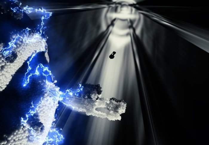
The technique involves vibrating the sample at ultrasonic frequencies so that the cell begins to fall apart. Electrical currents then applied an electric field to eject the protein machines out of the membrane and directly into a mass spectrometer – an instrument that can detect a molecule’s chemical ‘signature’, based on its mass.
Not only did the membrane protein machines survive the ejection; the analysis also revealed how they communicate with each other, are guided to their final location and transport their molecular cargo into the cell.
Professor Steve Matthews, from the Department of Life Sciences at Imperial, said: “With the development of this method, the application of mass spectrometry in biology will be taken to a new level, using it to make discoveries that would not have been possible before.”
Dr Sarah Rouse, also from the Department of Life Sciences at Imperial, said: “A longstanding question on the structure of one membrane machine from mitochondria has now been solved using this technique. Mitochondria are particularly interesting because there are several diseases that target them specifically, that we may now be able to design new therapies for.”
'
‘Protein assemblies ejected directly from native membranes yield complexes for mass spectrometry’ by Dror S. Chorev, Lindsay A. Baker, Di Wu, Victoria Beilsten-Edmands, Sarah L. Rouse, Tzviya Zeev-Ben-Mordehai, Chimari Jiko, Firdaus Samsudin5, Christoph Gerle, Syma Khalid, Alastair G. Stewart, Stephen J. Matthews, Kay Grünewald, and Carol V. Robinson is published in Science.
Article text (excluding photos or graphics) © Imperial College London.
Photos and graphics subject to third party copyright used with permission or © Imperial College London.
Reporter
Hayley Dunning
Communications Division