Nanoscale tweezers can perform single-molecule ‘biopsies’ on individual cells
Using electrical impulses, the ‘tweezers’ can extract single DNA, proteins and organelles from living cells without destroying them.
We are continuously expanding our knowledge on how cells function, but many unanswered questions remain. This is especially true for individual cells that are of the same type, such as brain, muscle or fat cells, but have very different compositions at the single-molecule level.
With our tweezers, we can extract the minimum number of molecules that we need from a cell in real time, without damaging it. Professor Joshua Edel
Cataloguing the diversity of seemingly identical cells can help researchers to better understand fundamental cellular processes and design improved models of disease, and even new patient-specific therapies.
However, traditional methods for studying these differences typically involve bursting the cell, resulting in all of its contents getting mixed. This results not only in the loss of spatial information – how the contents were laid out in relation to each other, but also in dynamic information, such as molecular changes in the cell over time.
Extraction without destruction
A new technique, developed by a team led by Professor Joshua Edel and Dr Alex Ivanov at Imperial College London, enables researchers to extract single molecules from live cells, without destroying them. The research, published today in the journal Nature Nanotechnology, could help scientists in building up a ‘human cell atlas’, providing new insights into how healthy cells function and what goes wrong in diseased cells.
Professor Joshua Edel, from the Department of Chemistry at Imperial, said: “With our tweezers, we can extract the minimum number of molecules that we need from a cell in real time, without damaging it.
"We have demonstrated that we can manipulate and extract several different parts from different regions of the cell – including mitochondria from the cell body, RNA from different locations in the cytoplasm and even DNA from the nucleus.”

Watch the video above for an explanation of the process and some microscopy footage.
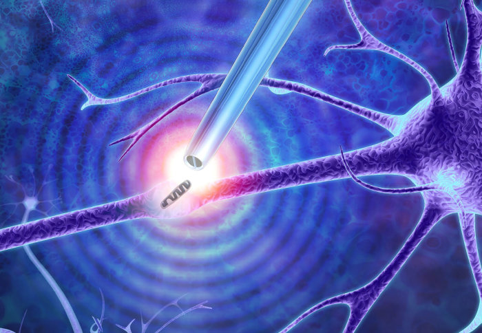
The tweezers are formed from a sharp glass rod terminating with a pair of electrodes made from a carbon-based material much like graphite. The tip is less than 50 nanometers (a nanometre is one-millionth of a millimetre) in diameter and is split into two electrodes, with a 10 to 20-nanometre gap between them.
By applying an alternating current voltage, this small gap creates a powerful highly localised electrical field that can trap and extract the small contents of cells such as DNA and transcription factors – molecules that can change the activity of genes.
Picking out individual molecules
The method is based on a phenomenon called dielectrophoresis. The tweezers generate a sufficiently high electric field enabling the trapping of certain objects such as single molecules and particles. The ability to pick out individual molecules form a cell sets it apart from alternative technologies.
These nanoscale tweezers could be a vital addition to the toolbox for manipulating single cells and their parts. Dr Alex Ivanov
The technique could potentially be used to carry out experiments not currently possible. For example, nerve cells require much energy to fire messages around the body, so they contain many mitochondria to help them function. However, by adding or removing mitochondria from individual nerve cells, researchers could better understand their role, particularly in neurodegenerative diseases.
Dr Alex Ivanov, from the Department of Chemistry at Imperial, said: “These nanoscale tweezers could be a vital addition to the toolbox for manipulating single cells and their parts. By studying living cells at the molecular level, we can extract individual molecules from the same location with unprecedented spatial resolution and over multiple points in time.
"This may provide a deeper understanding of cellular processes, and in establishing why cells from the same type can be very different to each other.”
Professor Edel added: “The whole project was only made possible by the unique know-how and abilities and enthusiasm of the young team members, including Dr Binoy Paulose Nadappuram and Dr Paolo Cadinu, amongst others, who all have diverse expertise and backgrounds.”
-
‘Nanoscale Tweezers for Single Cell Biopsies’ by Binoy Paulose Nadappuram, Paolo Cadinu, Avijit Barik, Alexander J. Ainscough, Michael J. Devine, Minkyung Kang, Jorge Gonzalez-Garcia, Josef T. Kittler, Keith R. Willison, Ramon Vilar, Paolo Actis, Beata Wojciak-Stothard, Sang-Hyun Oh, Aleksandar P. Ivanov, and Joshua B. Edel is published in Nature Nanotechnology.
Top image by Murray Robertson.
Article text (excluding photos or graphics) © Imperial College London.
Photos and graphics subject to third party copyright used with permission or © Imperial College London.
Reporter
Hayley Dunning
Communications Division