Cancer-causing HTLV-1 virus hijacks cellular machinery to establish infection
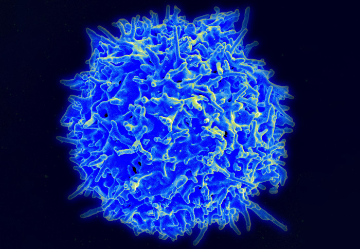
HTLV-1 targets T cells (pictured), a type of white blood cell / Credit: NIAID.
Scientists have used a cutting-edge visualisation technique to show how the human T-cell lymphotropic virus, type 1 (HTLV-1) infects immune cells.
HTLV-1 is a virus that affects T cells, a type of white blood cell which plays a crucial role in our immune system. Although most people infected with the virus do not experience symptoms, around two to five per cent will go on to develop adult T-cell leukaemia (ATL). Currently, between five and 20 million people worldwide are infected by HTLV-1 and no cure or treatment is available.
Now, a team of researchers at Imperial College London and the Francis Crick Institute have published new findings in Nature Communications showing in atomic detail how HTLV-1 infects immune cells. By providing a more nuanced understanding of how the virus establishes infection in the body, the research will help to support the development of new, targeted therapies.
Under the microscope
To shed light on how the virus infects cells, researchers led by Imperial’s Dr Goedele Maertens used a cutting-edge visualisation technique called cryogenic electron microscopy (cryo-EM). This allowed the team to take thousands of images of a specific molecule within the virus’ structure known as integrase.
In capturing these images, the researchers discovered that HTLV-1 uses integrase to hijack protein phosphatase 2A (PP2A), an important gatekeeping protein in immune cells. The virus is then able to insert a copy of itself into human DNA, representing an important tipping point in the infection cycle.
A step towards new therapies
Commenting on the wider significance of the findings, the study’s lead author Dr Michal Barski said: “This is an incredibly exciting time. 40 years after HTLV was discovered, we have finally laid the foundation for understanding with atomic precision how this virus integrates into human DNA and how it hijacks a key human protein to achieve this.”
Dr Goedele Maertens, senior author of the study and the head of Imperial’s HTLV laboratory, said: “This is a great step towards understanding how HTLV usurps the host cellular machinery to insert its genome into the host DNA.”
“This crucial moment in the replication cycle of the virus determines the fate of the patient: depending on where the viral DNA copy gets inserted, it could mean the patient will remain asymptomatic or develop a debilitating and sometimes fatal disease."
The new findings will support the design of a new drug which targets the integrase molecule and its interaction with immune cells.
‘Cryo-EM structure of the deltaretroviral intasome in complex with the PP2A regulatory subunit B56γ’ by Michal S. Barski et al. Is published in Nature Communications
Image: Scanning electron micrograph of a human T lymphocyte (also called a T cell) from the immune system of a healthy donor. Credit: NIAID.
Article text (excluding photos or graphics) © Imperial College London.
Photos and graphics subject to third party copyright used with permission or © Imperial College London.
Reporter
Ms Genevieve Timmins
Academic Services