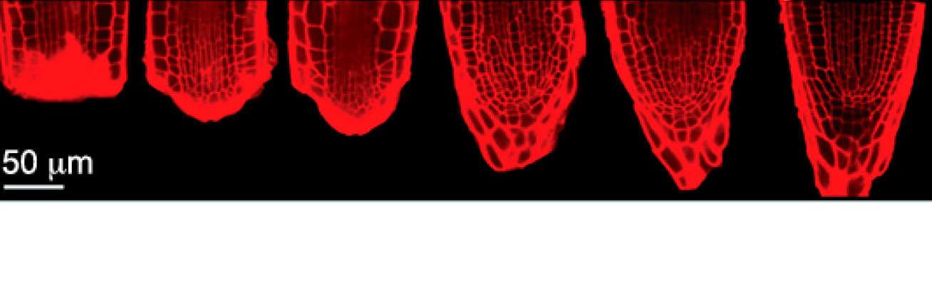
Root tip regeneration
We adopt root tip regeneration in Arabidopsis (Fig 1) as a model system to study plant tissue self-organisation. The long-term goal is to infer universal effective theories about the emergence of order in multicellular systems.
The main approach is to combine standard molecular genetics methods with novel live imaging setups and quantitative assays, to dissect the molecular and physical mechanisms regulating tissue self-organization.
Callus regeneration
A second model system used to address fundamental questions in plant tissue regeneration is callus, a mass of rather undifferentiated tissue that can be induced in vitro and is part of widely used microproagation methods.
Main questions
- Does the tissue reorganisation depend on well-defined organisers (instructive) or merely on local cell-cell interactions (true self-organisation)?
- What is the role of bioelectric patterns in tissue regeneration?
- What are the genetic, epigenetic and physical mechanisms regulating and constraining tissue regeneration in plants?
- How does organ regeneration differ from other examples of post-embryonic organogenesis?
- How can such morphological dynamics be quantitated and mathematically analysed?
- Can tissue self-organisation be controlled and enhanced in plants?
Light Sheet Fluorescence Microscopy
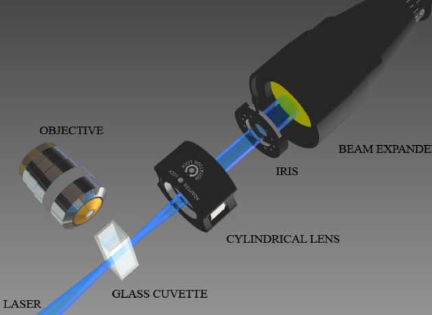
One of the tools used in this investigations is a unique setup based on light sheet microscopy, which allows us to acquire stacks of optical sections of growing and regenerating roots at cellular resolution (microns), with high temporal resolution (every 15 minutes) and for very long time (at least 7 days).
With this tool, we can observe in vivo the spatial and temporal dynamics of any fluorescent genetic reporter or molecular dye, during the entire duration of the regeneration process. The rich temporal series are used to test hypotheses on the fundamental mechanisms and to instruct ad hoc computational models.