3D ultrasound is an advanced imaging technique that can create three dimensional images by scanning an object using ultrasound from multiple directions and piecing together the various outputs into the final three dimensional image. Given the relative safety and cost effectiveness of conventional ultrasound, 3D ultrasound has the potential to offer significant capabilities in medical imaging that are yet to be realised. Currently, 3D ultrasound use in medicine is most commonly used as an elective procedure during pregnancy; very few uses outside of this exist in current clinical practice despite the [obvious] potential. Here at the College, one of the researchers working to change this is Dr Mengxing Tang. Through his research on ultrasound, Mengxing has been working to develop technologies that will allow 3D real time imaging of blood flow with high spatial and temporal resolution, potentially opening up a number of clinical areas in which such a technology could have an impact.
Understanding more about the ways in which blood flows around the body will be of great importance in improving the diagnosis and treatment of cardiovascular diseases such as atherosclerosis which causes hardening/narrowing of the arteries and strokes. For example, 3D ultrasound using contrast enhanced imaging will enable analysis of flow patterns through larger vessels and arteries and adverse pressure and shear on arterial walls, offering a valuable imaging tool for vascular diseases and potential biomarkers for their diagnosis and treatment monitoring. Furthermore, contrast enhanced ultrasound will also enable variations in the microcirculation structure to be studied which could provide long-term verification of improvement of damaged tissue, for example in the recovery of the heart muscle after a cardiac arrest or provide indications of potential issues in the upstream larger vessels and arteries.
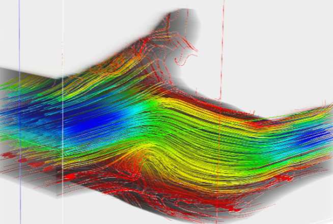
Through support from the EPSRC IAA, Mengxing and his researcher, Ms Virginie Papadopoulou have been developing a prototype system that is capable of the imaging and quantification of blood flow. Using a state-of-art contrast enhanced ultrasound research system (which you can read more about in Mengxing’s other pathways to impact project). By scanning in two different planes enables a full three dimensional picture to be built up potentially giving and unprecedented level of detail and information about the flow of blood through the cardiovascular system.
Ultimately the technology brings several other capabilities. The ultrasound system in this study takes images at several thousand frames per second (far quicker than the 20/second which the human eye can operate at) and as a result is able to track the quickly moving individual bubbles in the flow. Similarly to the way in which cars are monitored in fixed speed zones by average speed cameras on motorways, the system can determine the speed of micro-bubbles through a series of images collected. Due to its high imaging speed, the technology also has the potential to offer much improved imaging of a beating heart than current available clinical techniques.