The ATR-FTIR imaging enables one to make spatially resolved chemical snapshots of microscopic objects, specifically cells and tissue, as a function of time ("chemical photography") and this could be fully exploited in the study of biological objects such as cells and tissues (e.g. see Kazarian S. G., Chan K. L. A. "Applications of ATR-FTIR spectroscopic imaging to biomedical samples" Biochimica et Biophysica Acta (BBA) - Biomembranes 1758 (2006) 858-867 or Kazarian S. G., Chan K. L.A. "ATR-FTIR spectroscopic imaging: recent advances and applications to biological systems" (Tutorial review) Analyst 138 (2013) 1940-1951 (doi) or our recent invited review: Chan K. L. A., Kazarian S. G. "Attenuated total reflection Fourier-transform infrared (ATR-FTIR) imaging of tissues and live cells" Chemical Society Reviews (2016) 45, 1850-1864 (doi), which has featured in the Department web-site, see also the most recent Trends article by Kimber J. A., Kazarian S. G. "Spectroscopic imaging of biomaterials and biological systems with FTIR microscopy or with quantum cascade lasers" Analytical and Bioanalytical Chemistry (2017) 409 (25) 5813-5820 which is Open Access (doi) and featured on the cover of October issue 2017. Most recently we have demonstrated for first time an application of micro ATR-FTIR spectroscopic imaging with variable angle of incidence to obtain a depth profile of tissue samples. This novel investigation reveals a new analytical insight into studying the variation of tissue composition with depth in a non-destructive manner and provides improved cancer diagnostic, Analyst (2019) (doi). Diagnostic applications with machine learning wererecently developed in FTIR spectroscopic imaging of colon tissues, Anal Bioanal Chem (2019) (doi) OPEN ACCESS. Please see our recent Perspective article for our outlook in this field.
The range of our applications of FTIR spectroscopic imaging to biomedical field is broad. These were achieved by collaboration with scientists in different biomedical disciplines. Our successful collaboration with experts in atherosclerosis, Prof. M. J. Lever and Prof. Weinberg from the Department of Bioengineering at Imperial College London and Prof. R. K. Upmacis from Department of Pathology and Laboratory Medicine, Center of Vascular Biology at Cornell University showed ability of ATR-FTIR imaging to analyse arterial samples; while collaboration with the Department of Materials demonstrated success of imaging for biomaterials in tissue engineering. Collaboration with Dr. Mantalaris (imperial College London) resulted in successful applications of ATR-FTIR imaging in stem cells research while collaboration with Prof. N. Chayen (Department of Bio-Molecular Medicine, Imperial College London) led to the first example of imaging of crystallization of proteins. This research currently continues in collaboration with Dr. Bernadette Byrne (Department of Life Sciences, Imperial College London) via current grant from BBSRC (BRIC) "Application of ATR-FTIR imaging to industrial scale production of therapeutic antibodies". These successful applications open a broad and exciting range of opportunities for medical research. Indeed, ATR-FTIR spectroscopic imaging is destined to be successful in biomedical research and analytical applications.
Projects in this field have included: 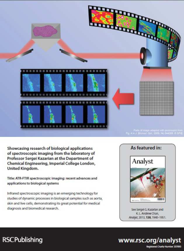
Protein crystallisation and aggregation
Skin and transdermal drug delivery