Tabs 1
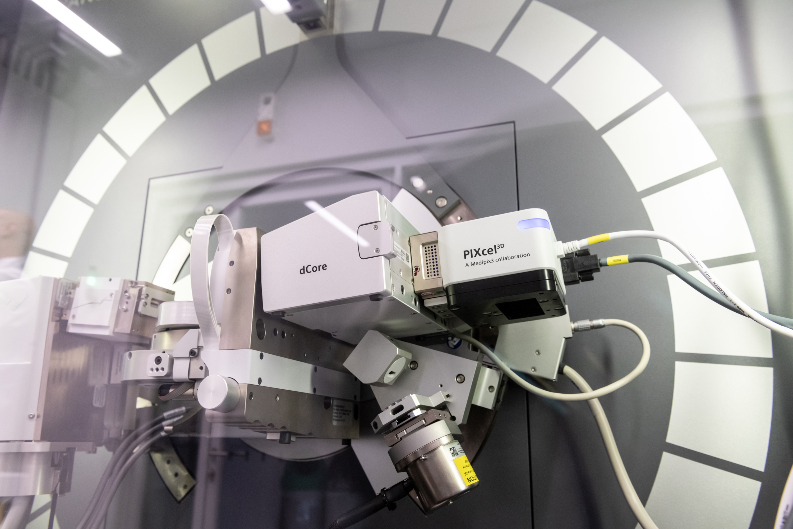 Model: Malvern Analytical Empyrean MultiCore High-Performance X-ray Diffractometer
Empyrean covers the most extensive X-ray diffraction, scattering and imaging applications in one instrument. The MultiCore Optics enable the widest variety of measurements without any manual intervention. Empyrean can measure all sample types - from powders to thin films, from nanomaterials to solid objects - on a single instrument.
Moreover, Empyrean not only meets the high expectations of scientists and XRD experts today but will continue to do so as research themes evolve. Empyrean is ideal for teaching purposes, thanks to the large doors that open entirely, allowing access to the system to several people, but at the same time is perfect for performing measurements in demanding R&D environments in several industries.
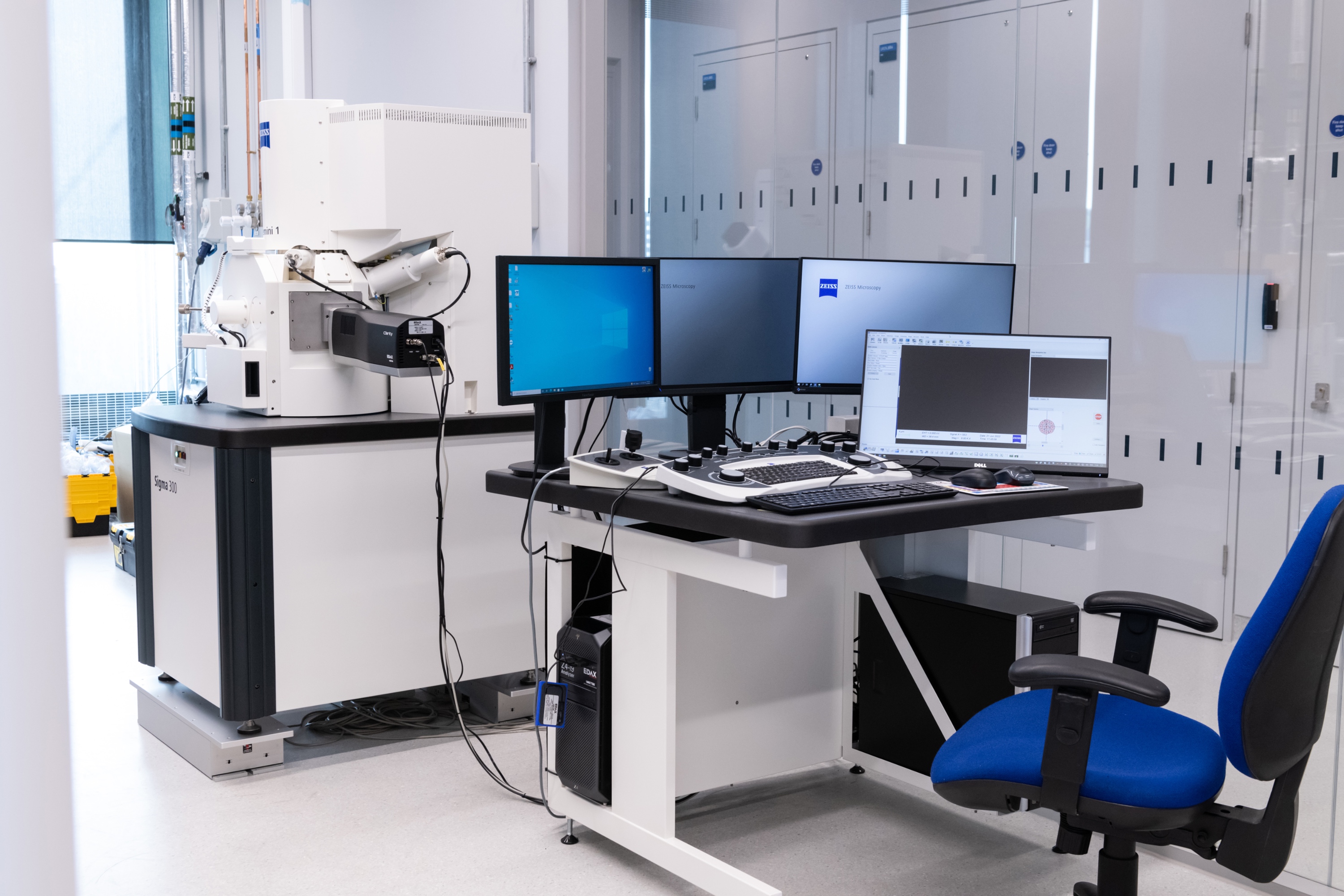
Model: Gemini 1 Zeiss Sigma 300 w/ Clarity EBSD
Combine field emission SEM (FE-SEM) technology with analytics. Profit from proven Gemini electron optics. Choose from a variety of detector options: image particles, surfaces, and nanostructures in materials science investigate semiconductor or medical devices, and geological or biological samples.
Save time with the semi-automated 4-step workflow of Sigma: structure your imaging and analysis routines and increase productivity. FE-SEM users of all disciplines in research and industry labs now benefit from a resolution of 1.3 nm at 1 kV in ZEISS Sigma 500 and better usability.
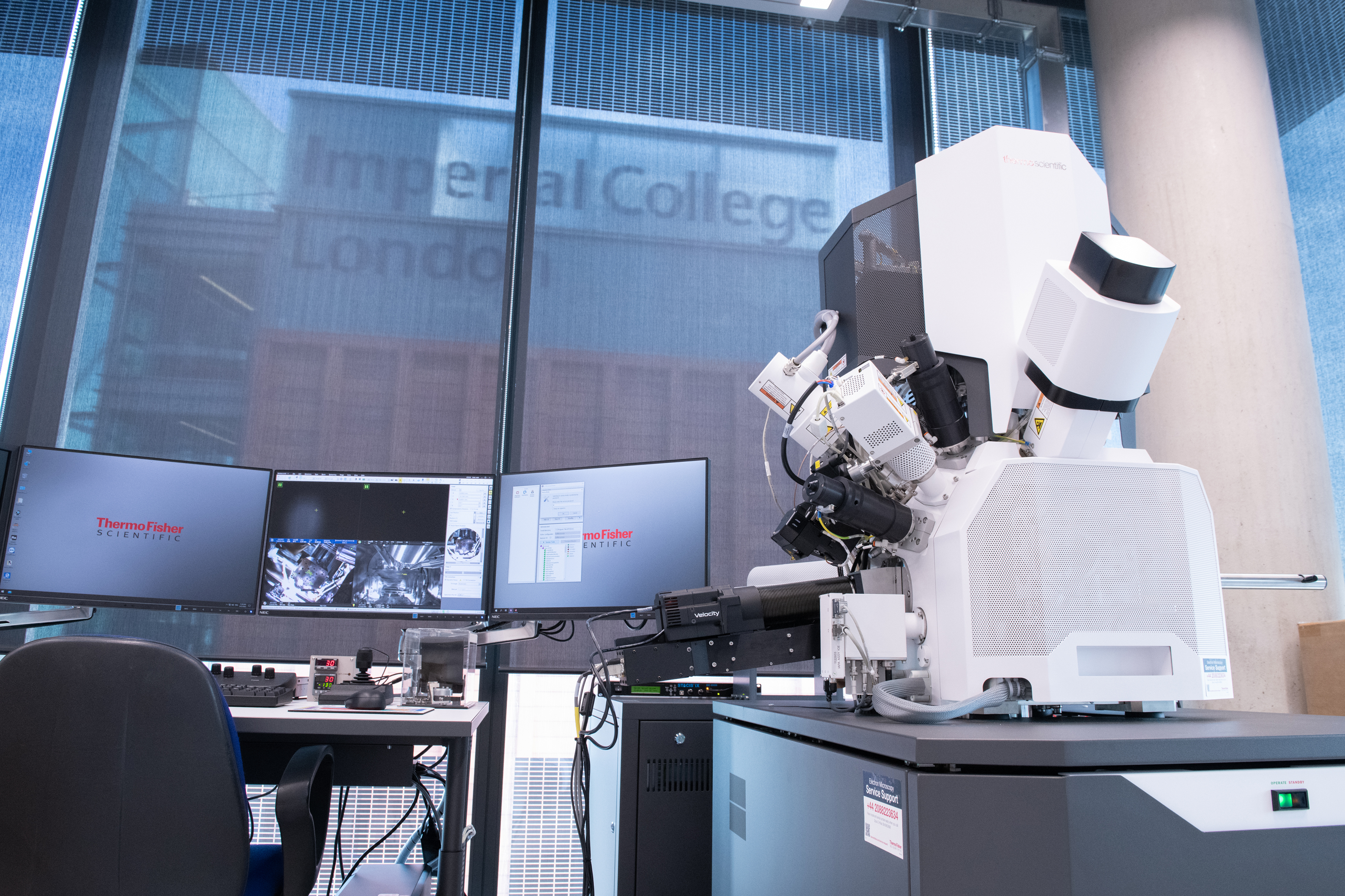
Model: Thermo Scientific Scios - 2 HiVac with Retractable RGB CL Detector w/ Velocity EDSB
Backscattered electron and secondary electron imaging
The innovative NICol electron column provides the foundation of the system’s high-resolution imaging and detection capabilities. It offers excellent nanoscale details, with a wide range of working conditions, whether operating at 30 keV in STEM mode (to access structural information) or at lower energies (to obtain charge-free, detailed surface information).
With its unique in-lens Thermo Scientific Trinity Detection System, the Scios 2 DualBeam is designed to simultaneously acquire angular and energy-selective secondary electron (SE) and BSE imaging. Fast access to detailed nanoscale information is possible not only top-down but also on tilted specimens or cross-sections.
Optional below-the-lens detectors and an electron-beam-deceleration mode ensure quick and easy simultaneous collection of all signals, revealing the minor features in a material surface or cross-section. Fast, accurate, and reproducible results are obtained thanks to the unique NICol column design with full auto alignments.
The FIB protocols we can offer external users comprise Imaging, Patterning, Deposition and Cross-sectioning. If individual users are experienced in other techniques, we may be able to accommodate these too. If this is the case, please get in touch with your level of experience in the desired technique, so we can assess your project and discuss potentially accommodating your needs.
How to access
To access our facilities, please email royce@imperial.ac.uk.
.jpg)