What is the Multi-‘omics Atlas Project?
We use:
- Well-phenotyped tissues (AD vs non-disease control)
- Open and integrated datasets, all linked to the brain and region from which they were derived
- Standardised data acquisitions and data analysis pipeline
- Multiple platforms (e.g., imaging mass cytometry, genomics, transcriptomics, epigenomics, metabolomics, proteomics)
- Data from studies of the same brains and regions by other investigators as the project evolves
Project background
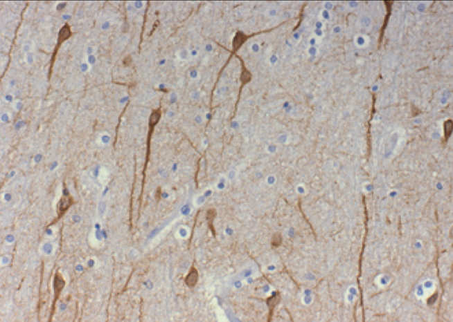
Neurodegenerative disease manifests through interactions between cells throughout the brain; it is not a cell- or region- autonomous process and the pathology evolves in a complex way from primary dysfunction (accumulation of misfolded proteins) through to secondary pathology (inflammation and synapse loss). Characterising these pathological changes is central to the development of new diagnostic, monitoring or therapeutic tools. There is an immediate opportunity to rapidly improve understanding of these processes by exploitation of recent, transformative advances in multi-omics, imaging and computational methods.
Overview
Working with Brains for Dementia Research, the UK Brain Banks Network and Alzheimer Scotland Dementia Research Centre, our team has developed a tissue resource of carefully selected, well-annotated and centrally managed brains with one hemisphere frozen and the other hemisphere fixed for microscopic analysis. Material from standardised sampling regions are studied, both at the level of the single cell and by observing multi-cellular characteristics. As we established the resource, we tackled other key, previously unanswered questions about tissue quality to help refine similar studies in future. This will include how time between death and freezing of tissue impacts its quality and what is the best measure of tissue quality.
All data and spatial information are compared across different brain regions and at different stages of the disease. By studying tissue at the early stage of illness, we determine the most optimal time to target the disease before widespread neurodegeneration takes place. Efforts to target the late stages of Alzheimer’s disease have thus far proved ineffective, and the field is now focusing efforts on developing therapies to be given as early as possible to alter the course of the illness.
During the initial three-year pilot, we studied 14 brains of human donors who had Alzheimer’s at different stages of the disease, and 14 brains of donors without Alzheimer's Disease. The project was designed to be extended to include new techniques as they are developed, using the same set of donor brains, and to add new data to the existing dataset. We have also expanded the study to include cases from genetically-stratified cohorts. We hope that the study could eventually be expanded to map the pathology of other neurodegenerative diseases.
![]()
