Connectomics is the production and study of comprehensive maps of an organism’s nervous system. It uses high-throughput imaging and histological procedures to map neural connections – the underlying premise being that, to understand (and repair) a circuit, it is necessary to obtain its “wiring diagram”. Connectomic studies of cortical tissue show substantial promise to make advances in understanding brain disorders such as dementia, a major translational focus of the Centre for Neurotechnology.
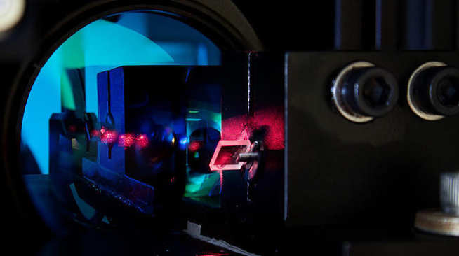
Thanks to equipment funds from the EPSRC, together with contributions from a number of research laboratories at Imperial, the Centre for Neurotechnology has established a cellular resolution Connectomics Facility based on a TissueVision TissueCyte1000 serial two photon tomography platform. This platform allows axon tracing to be performed, with single cell resolution, in whole mouse brains, or large sections of a human brain. It is an automated system capable of two-photon, sub-micron resolution imaging of entire small organs, ex vivo. A mouse brain, or similarly sized organ, can be serially imaged over several days to generate three-dimensional data that can be used to map structures of interest such as cells, plaques or white/grey matter morphology.
The facility is open for use by any member of Imperial College.
Booking is via the PPMS online system. Please see our page for further details.
For enquiries about the facility, please contact Nawal Zabouri.

Useful links
Funding support
We acknowledge the support of the EPSRC and Wellcome Trust in funding research facilities for the Centre for Neurotechnology.


