Contact
Dr Mitch Chen
mitchell.chen@imperial.ac.uk
What we do
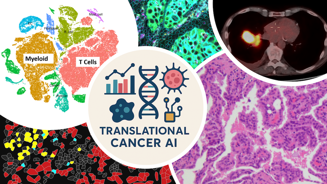
We use artificial intelligence, medical imaging and advanced spatial molecular profiling to discover explainable biomarkers for precision cancer medicine, with a focus on lung cancer.
We integrate medical imaging (CT and 18F-FDG PET) with spatial biological profiling data, including RNA, protein and histology, to understand tumour biology and predict treatment response, toxicity and patient outcomes.
Why it is important
Cancer treatment is becoming increasingly personalised, yet selecting the right treatment for the right patient remains challenging. Patients with radiologically and histologically similar cancers can respond very differently to the same therapy, with some experiencing serious treatment-related adverse events without meaningful clinical benefit.
Medical imaging contains rich quantitative information that is not readily captured by conventional visual assessment. AI can unlock this information non-invasively and at scale, without requiring additional patient tests or substantially increasing clinical workload.
How it can benefit patients
We develop novel imaging and molecular biomarkers that help identifying patients who are most likely to benefit from a treatment. Rather than replacing established biomarkers, our tools are designed to complement them, making cancer treatment more precise, effective and safe for individual patients.
Summary of current research
Our research spans five complementary areas:
Current research
- 1) Explainable AI biomarkers for precision cancer treatment
- 2) Linking imaging with tumour biology
- 3) Foundation and world models for cancer imaging AI
- 4) Body composition and sarcopenia
- 5) Translating AI into clinical practice
We develop imaging and molecular biomarkers within an explainable and causal framework, to predict cancer treatment response, survival and toxicity.
Through IMPACT, we integrate medical imaging with spatial molecular profiling to uncover relationships between imaging phenotypes, the tumour–immune microenvironment and treatment response, supporting biologically grounded biomarkers for treatment selection.
We develop foundation models for generalisable cancer imaging analysis, and world models that integrate imaging and biological data to model tumour behaviour and investigate mechanisms of cancer progression and treatment response.
Through ASCEND, we use automated CT analysis for opportunistic screening of sarcopenia, to investigate how skeletal muscle and other body-composition measures relate to treatment tolerance, toxicity and outcomes across cancers.
We validate AI tools across independent, multicentre datasets, with the goal of translating our discoveries into routine clinical practice.
Additional information
- IMPACT (REC: 25/HRA/4804): Imaging and Molecular Profiling and Integration for Precision Cancer Treatment (Oct 2025 – Present)
- ASCEND (REC: TBC): Automatic Sarcopenia CT Evaluation Network & Data (Oct 2026 – )
- AID-CXR (REC: 24/HRA/4895): Assessing artificial Intelligence for the Diagnosis of Cancer on chest-XRays (Nov 2024 – Present)
- Anifowose JO, Li Z, Agarwal G, Aboagye EO, O'Regan DP, Ariff B, Copley SJ, Chen M. Automated opportunistic cardiovascular risk assessment in non-small cell lung cancer patients on routine chest CT using an optimised nnU-net framework. BMC Med Imaging. 2026 Mar 3;26(1):179. doi: 10.1186/s12880-026-02252-z. PMID: 41772492; PMCID: PMC13064248.
- Chen M, Copley SJ, Linton-Reid K, Viola P, Han Y, Cortellini A, Lu H, Mani A, Bahket M, Pinato DJ, Power D, Rockall AG, Aboagye EO. A radio-genomics biomarker for precision epidermal growth factor receptor mutation targeting therapy in non-small cell lung cancer. Sci Rep. 2026 Mar 6;16(1):12416. doi: 10.1038/s41598-026-42948-4. PMID: 41792401; PMCID: PMC13083983.
- Chen M, Copley SJ, Han Y, Arshad MA, Viola P, Linton-Reid K, Stoycheva T, Cook GJR, Landau D, Chua S, O'Connor R, Dickson J, Power D, Rockall AG, Barwick TD, Aboagye EO. An explainable imaging-clinical biomarker for non-small cell lung cancer prognostication based on normalised hotspot to centroid distance and [18F]FDG PET/CT radiomics. Eur J Nucl Med Mol Imaging. 2026 Apr;53(5):3195-3212. doi: 10.1007/s00259-025-07659-4. Epub 2025 Dec 12. PMID: 41381759; PMCID: PMC13013418.
- Chen M, Linton-Reid K, Aboagye EO, Copley SJ. Translating radiomics into clinical practice: A step-by-step guide to study design and evaluation. Clin Radiol. 2025 Nov;90:107053. doi: 10.1016/j.crad.2025.107053. Epub 2025 Aug 22. PMID: 40962646.
- Chen M, Lu H, Copley SJ, Han Y, Logan A, Viola P, Cortellini A, Pinato DJ, Power D, Aboagye EO. A Novel Radiogenomics Biomarker for Predicting Treatment Response and Pneumotoxicity From Programmed Cell Death Protein or Ligand-1 Inhibition Immunotherapy in NSCLC. J Thorac Oncol. 2023 Jun;18(6):718-730. doi: 10.1016/j.jtho.2023.01.089. Epub 2023 Feb 10. PMID: 36773776.
- Chen M, Copley SJ, Viola P, Lu H, Aboagye EO. Radiomics and artificial intelligence for precision medicine in lung cancer treatment. Semin Cancer Biol. 2023 Aug;93:97-113. doi: 10.1016/j.semcancer.2023.05.004. Epub 2023 May 19. PMID: 37211292.
- Chen M, Helm E, Joshi N, Gleeson F, Brady M. Computer-aided volumetric assessment of malignant pleural mesothelioma on CT using a random walk-based method. Int J Comput Assist Radiol Surg. 2017 Apr;12(4):529-538. doi: 10.1007/s11548-016-1511-3. Epub 2016 Dec 27. PMID: 28028655; PMCID: PMC5362666.
Our researchers
Dr Mitch Chen
/prod01/channel_3/media/images/people-list/Mitch-Chen.jpg)
Dr Mitch Chen
MRC Clinician Scientist
Jillian Sieh
/prod01/channel_3/media/images/people-list/Jillian-Sieh.jpeg)
Jillian Sieh
Research Associate
Meng Zhou
/prod01/channel_3/media/images/people-list/Meng-Zhou.jpeg)
Meng Zhou
Research Associate
Charlie Johnson
/prod01/channel_3/media/images/people-list/Charlie-Johnson.jpeg)
Charlie Johnson
Research postgraduate
Ethan Chang
/prod01/channel_3/media/images/people-list/Ethan-Chang.jpeg)
Ethan Chang
Research Postgraduate
Pranathi Ratvikumar
/prod01/channel_3/media/images/people-list/Pranathi-Ratvikumar.jpeg)
Pranathi Ratvikumar
Research Postgraduate
Ainkaran Santhirasekaram
/prod01/channel_3/media/images/people-list/Ainkaran-Santhirasekaram.png)
Ainkaran Santhirasekaram
Clinical Lecturer
Girija Agarwal
/prod01/channel_3/media/images/people-list/Girija-Agarwal.jpeg)
Girija Agarwal
NHS Clinical AI Fellow
