MRI
- Magnetic Resonance Imaging Duodenoscope
- Comparison of MR Endoscopy and Endoscopic Ultrasound
- Micro-coils for Local MR-Thermometry During Laser Liver Ablation
- Design of Magneto-Inductive Magnetic Resonance Imaging Catheters
- Magneto-Inductive Catheter Receiver for Magnetic Resonance Imaging
- Frequency Scaling of Catheter-Based Magneto-Inductive MR Imaging Detectors
- Metamaterial Magnetic Resonance Imaging Duodenoscope
- Surgical Wound Monitoring by MRI with a Metamaterial-based Implanted Local Coil
Richard Syms, Ian Young, Chris Wadsworth, Simon Taylor-Robinson, Marc rea
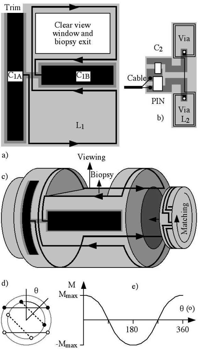
We have demonstrated a side-viewing duodenoscope capable of both optical and magnetic resonance imaging (MRI). The instrument is constructed from MR-compatible materials, and combines a coherent fibre bundle for optical imaging, an irrigation channel and a side-opening biopsy channel for the passage of catheter tools with a tip saddle coil for radio-frequency signal reception. The receive coil is magnetically coupled to an internal pickup coil to provide intrinsic safety. Impedance matching is achieved using a mechanically variable mutual inductance, and active decoupling by PIN-diode switching. 1H magnetic resonance imaging of phantoms and ex vivo porcine liver specimens was carried out at 1.5 T. An MRI field-of-view appropriate for use during endoscopic retrograde cholangiopancreatography (ERCP) was obtained, with limited artefacts, and a signal-to-noise ratio advantage over a surface array coil was demonstrated.
 |
|---|
| a) external coil, b) internal coil, c) integration on duodenoscope, d) variable transformer, e) variation of mutual inductance with angle. |
 |
|---|
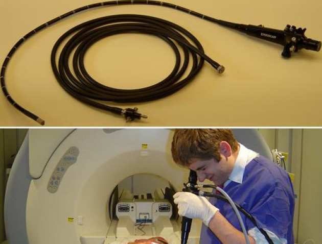
| Completed instrument and umbilical (upper); clinical evaluation in the magnet room (lower). |
 |
|---|
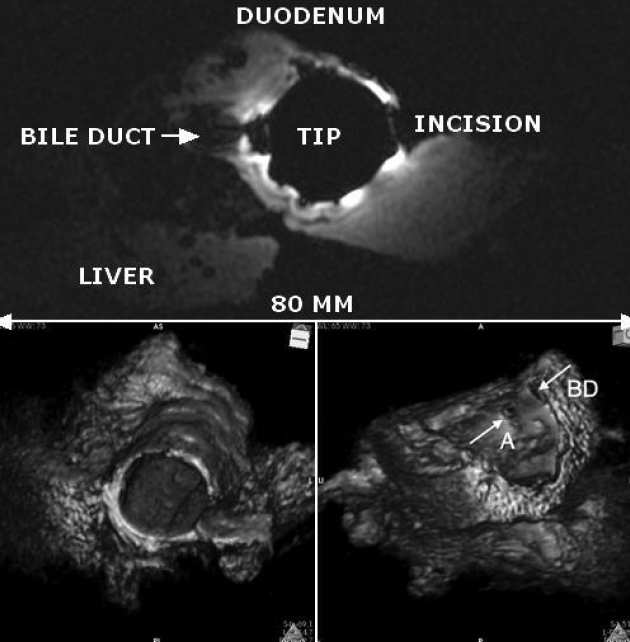
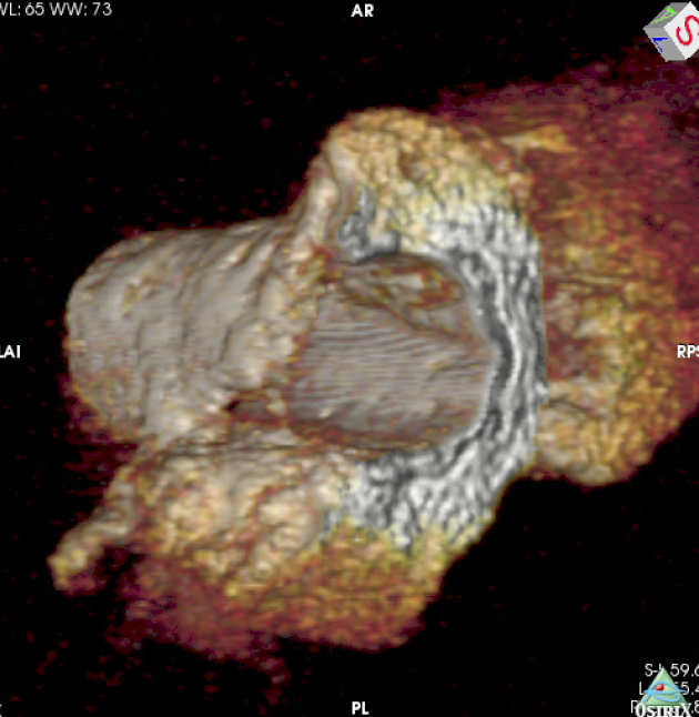
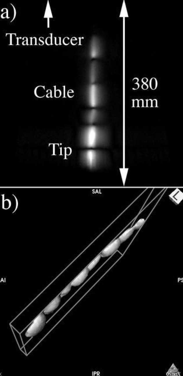
| Axial slice image of the porcine liver specimen obtained using the duodenoscope receiver (upper); 3D reconstructions from axial slices (lower). |
Richard Syms, Chris Wadsworth, Ian Young, Marc Rea and Simon Taylor-Robinson
Endoscopic ultrasound (EUS) is a well-established, safe modality for real-time imaging of the duodenum and nearby ductal systems. However, all echo-ranging imaging suffers from artifacts, the most significant being reverberation between impedance discontinuities. As a result, concentric artifacts dominate the near field even when a balloon is used. Acoustic shadowing is caused by strongly reflecting structures (voids such as lungs and bowel; bone and gallstones), while enhancement is caused by weakly attenuating fluid-filled structures (bladder and cysts). These artifacts cannot be suppressed without eliminating the fundamental contrast mechanism, unless a nonlinear contrast agent is used. As a result, EUS images are patient and operator dependent, and often hard to interpret. Although slower, internal MRI may therefore offer advantages, and suitable gastroscopes have already been developed. We have compared the performance of a new MR-imaging duodenoscope with EUS, and find significant advantages in contrast, resolution and reduction of artefacts.
 |
|---|
 |
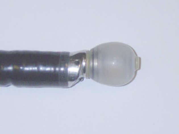
| Tips of ultrasonic (upper) and MR-imaging (lower) duodenoscopes. |
 |
|---|
| Ultrasound image of porcine duodenum and bile duct. |
 |
|---|
| 3D MR image of porcine duodenum and bile duct, reconstructed from axial slices. |
Evi Kardoulaki, Richard Syms, Ian Young, Marc Rea and Wady Gedroy
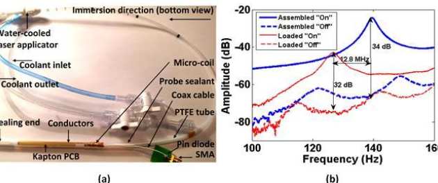
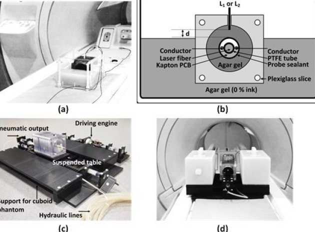
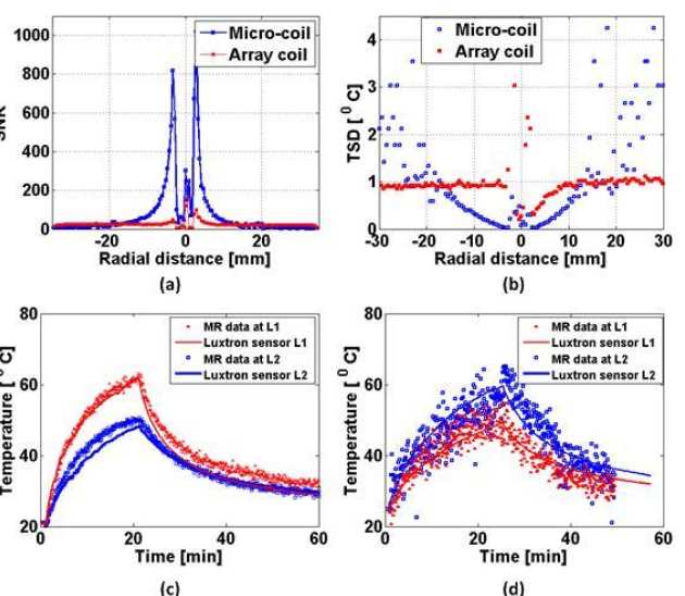
We have investigated whether local flexible micro-coils integrated with ablation catheters can improve the temperature accuracy during MR-thermometry in liver laser interstitial thermal therapies (LITTs). A liver-mimicking gel phantom was used to assess micro-coil derived image quality and sensitivity and ensure such coils can provide adequate FOV and resolution for the target application at 3T. The impact of liver motion was assessed using a MR-compatible hydraulic motion simulator. The thermal profile of a static phantom during an Nd:YAG laser ablation was monitored using reference-based PRF MR-thermometry and the robustness of the method under respiratory gating was evaluated on an un-heated phantom. The results were compared with the best locally available array coil for LITTs. Micro-coils improve the temperature accuracy by 1.5-10 times in a radius matching typical lesion dimensions and enable 1 mm image resolution. The resolution can be maintained during motion by using short acquisition time sequences while the SNR remains sufficient for accurate MR-thermometry. The temperature error on the un-heated phantom under respiratory gating does not exceed 1oC.
 |
|---|
| a) Modified laser applicator with integrated micro-coil receiver and (b) S-parameters of the micro-coil tuned and matched for use in a 3T scanner. |
 |
|---|
| a) Arrangement for the static SNR and MR-thermometry comparison studies, (b) schematic of an axial thermometry slice, c) hydraulic simulator of liver motion due to breathing and (d) complete arrangement for the assessment of motion artefacts. |
 |
|---|
| a) Radial SNR profile of the thermometry baseline image, b) respective radial temperature standard deviation; c) and d) Comparison of MR-inferred transient temperatures with the fluoro-optic readings. |
Khoonsake Segkhoonthod, Richard Syms and Ian R. Young
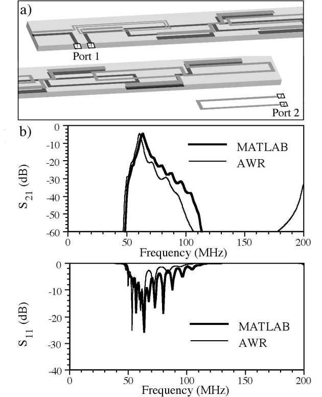
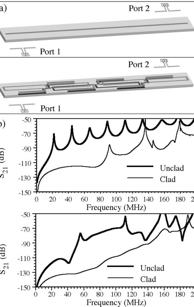
We have modeled a catheter-based RF receiver for internal magnetic resonance imaging. The device consists of a double-sided thin-film circuit, mounted on a hollow catheter. The system was originally designed for biliary ductal imaging, but is also potentially useful for vascular imaging. Signals are detected using a resonant L-C circuit at the catheter tip, transmitted along the catheter using an array of coupled L-C resonators, and coupled into a conventional RF system using a demountable inductive transducer. Protection against external B1 and E fields is obtained by using figure-of-eight-shaped elements with an electrical length shorter than that of an immersed half-wave dipole. Electromagnetic modeling software (AWR Microwave Office) has been used to analyze a system designed for 1H imaging at 1.5 T, determine the effect of the tissue surround, demonstrate signal detection and transmission and verify intrinsic safety.
 |
|---|
| Thin-film detector: a) and b) circuit, and c) integration on catheter. |
 |
|---|
| Detector simulation: a) AWR model; b) frequency variations of S11 and S21 as predicted by AWR and MATLAB. |
 |
|---|
| Simulation of electric decoupling: a) AWR models and b) frequency variations of S21 for wire and cable, with and without cladding. |
Richard Syms, ian Young, Munir Ahmad, Simon Taylor-Robinson and Marc Rea
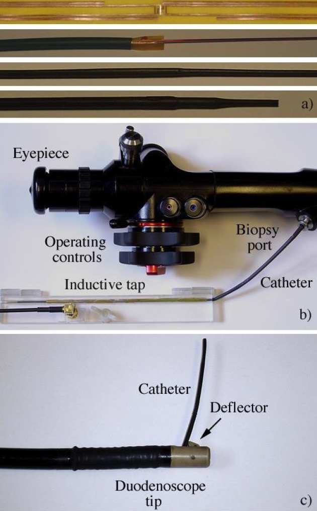
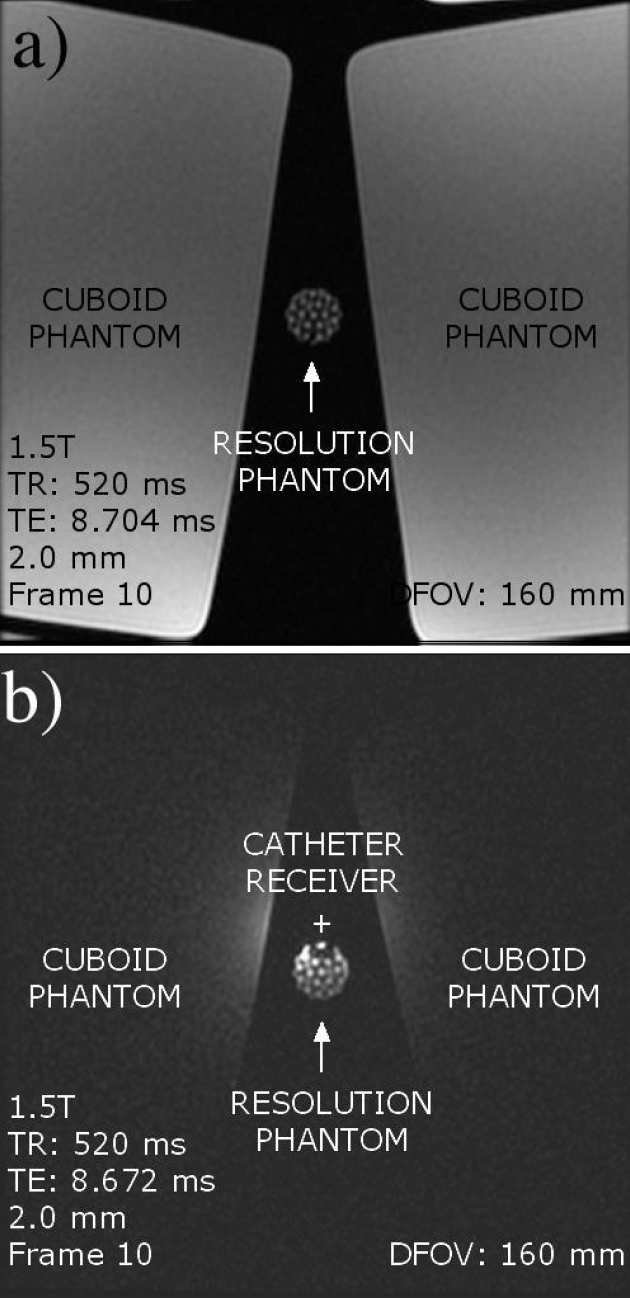
We have demonstrated a catheter-based RF receiver for internal magnetic resonance imaging is demonstrated. The device consists of a double-sided thin-film circuit, wrapped around a hollow catheter and sealed in place with heatshrink tubing. Signals are detected using a resonant L-C circuit at the catheter tip, and transmitted along the catheter using an array of coupled L-C circuits arranged as a magneto-inductive waveguide, a form of low frequency metamaterial. Coupling to a conventional RF system is accomplished using a demountable inductive transducer. Protection against external B1 and E fields is obtained by using figure-of-eight elements with an electrical length shorter than that of an immersed dipole. The system is primarily designed for biliary imaging, can pass the biopsy channel of a side-opening duodenoscope and is guidewire-compatible, potentially allowing clinicians to implement MR image guided procedures without changing their standard practice. Decoupling against B1 and E fields has been verified, and in vitro 1H magnetic resonance imaging with sub-mm resolution demonstrated at 1.5 T using phantoms.
 |
|---|
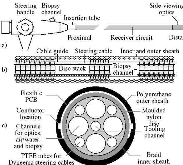
| a) Stages in construction of catheter-based receiver; b) receiver with demountable transducer attached and passing into the biopsy channel of a non-magnetic duodenoscope; c) receiver passing from the side-port. |
 |
|---|
| Axial 1H MR images obtained using a) an 8-element array and b) the catheter receiver; c) high-resolution image obtained using the catheter. |
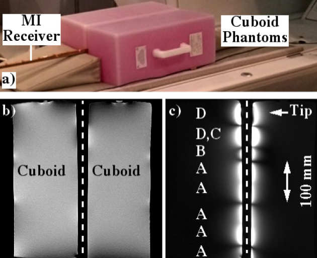
Richard Syms, Ian Young, Marc Rea
We have developed frequency-scaling rules for catheter-based magneto-inductive magnetic resonance imaging detectors, intended for in vivo imaging of the vascular and biliary ductal systems. The design is based on a cascade of magnetically coupled L-C resonators, fabricated as a thin-film circuit and mounted on a catheter. Intrinsic safety is introduced using resonant elements designed to avoid coupling to uniform RF magnetic and electric fields. We have used these rules to demonstrate frequency scaling of developed designs from 1.5 T to 3 T, and carried out mapping of reception patterns and high-resolution 1H imaging in a 3 T clinical scanner.
 |
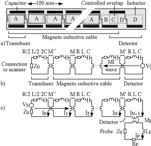
|---|
| CAD layout, PCB, completed catheter and inductive coupling transducer for a magneto-inductive catheter receiver. |
 |
|---|
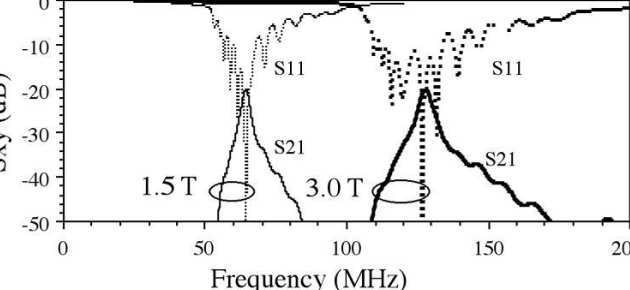
| Frequency response of catheter receivers designed for operation at 1.5T and 3 T. |
 |
|---|
| Coronal image of cuboid phantom obtained with an MI catheter receiver, and reception pattern reconstructed from coronal images. |
Richard Syms, Evi Kardoulaki and Ian Young
Simon Taylor-Robinson, Chris Wadsworth and Marc Rea (St Mary’s Hospital)
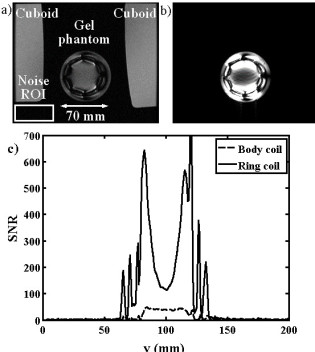
We have developed a magnetic resonance imaging duodenoscope, by combining non-magnetic endoscope components with a thin-film receiver based on a magneto-inductive waveguide. The waveguide elements consist of figure-of-eight shaped inductors formed on either side of a flexible substrate and parallel plate capacitors that use the substrate as a dielectric. Operation has been simulated using equivalent circuit models and by computation of sensitivity patterns. Circuits have been fabricated for operation at 127.7 MHz by double-sided patterning of copper-clad Kapton and assembled onto non-magnetic flexible endoscope insertion tubes. Operation has been verified by bench testing and by 1H MRI at 3T using phantoms. The receiver can form a segmented coaxial image along the length of the endoscope, even when bent, and shows a signal-to-noise-ratio advantage over a surface array coil up to three times the tube diameter at the tip. Initial immersion imaging experiments have been carried out and confirm an encouraging lack of sensitivity to RF heating.
 |
|---|
| Mechanical arrangement of metameterial endoscope. |
 |
|---|
| Electrical layout of thin film PCB (a), and equivalent circuit models for imaging (b) and electrical testing (c). |
 |
|---|
| Arrangement for magnetic resonance imaging with cuboid phantoms (a), body coil image showing effective decoupling (b) and metamaterial coil image, showing segmented field of view (c). |
Hanan Kamel, Richard Syms, Evi Kardoulaki, Marc Rea
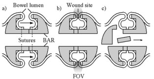
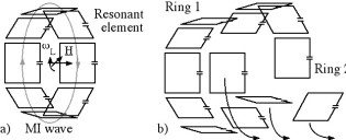
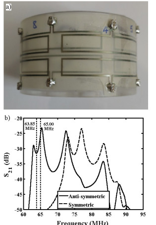
We have demonstrated an early prototype implantable sensor for monitoring surgical wounds after bowel reconstruction. The sensor consists of a coupled pair of 8-element magneto-inductive ring resonators, designed for mounting on a biofragmentable anastomosis ring to give a local increase in signal-to-noise ratio near an annular wound during 1H magnetic resonance imaging. Operation on an anti-symmetric spatial mode is used to avoid coupling to the B1 field during excitation, and a single wired connection is used for MRI signal output. The electrical response and field-of-view have been estimated theoretically. Prototypes have been constructed from flexible elements designed for operation at 1.5 T, electrical responses have been characterized and local SNR enhancement has been confirmed using agar gel phantoms.
 |
|---|
| Operation of a biofragmentable anastomosis ring: a) implantation during surgery, b) wound healing and c) breakdown |
 |
|---|
| Electrical arrangement of a) single and b) double magneto-inductive ring resonators. |
 |
|---|
| Prototype sensor: a) arrangement of thin film circuits before encapsulation; b) mode spectrum obtained by inductive probing. |
 |
|---|
| Results of Magnetic resonance imaging at 1.5 T: axial slice images obtained using a) the body coil and b) the sensor; c) variation of SNR with vertical position obtained using the two coils. |
Optical monitoring
- Real time optical monitoring of RF small bowel tissue fusion
- Refractive index of human haemoglobin in the optical range
- Kramers-Kronig analysis of the refractive index of human haemoglobin
Timmy Floume, Richard Syms, George Hanna, Ara Darzi (St Mary’s Hospital)
 |
|---|
| Lab-based optical measurement of RF tissue fusion |
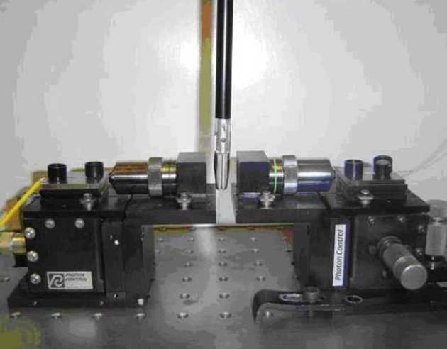
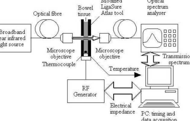
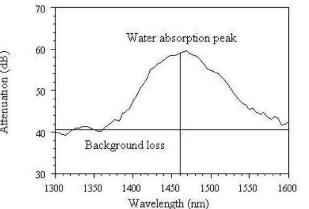
Radio frequency (RF) tissue fusion is a novel method of tissue approximation that can seal tissue without the need for sutures or staples, based on tissue transformations induced by Joule heating and mechanical pressure. RF delivery must be controlled and optimised to obtain a reproducible, reliable seal. At present, tissue impedance is used for feedback during RF blood vessel sealing. We have developed a method of real-time optical measurement to improve understanding of the tissue modifications induced by RF fusion. Near infrared transmission spectroscopy has been used for dynamic acquisition of the tissue attenuation spectrum, and the modified Beer-Lambert law has been used to extract optical parameters. Simultaneous temperature measurement shows that tissue coagulation alters scattering losses and that tissue dehydration induces changes in the water absorption band near 1450 nm wavelength.
 |
|---|
| Experimental arrangement |
 |
|---|
| Water absorption band in pig bowel tissue |
O. Sydoruk (in collaboration with Erlangen-Nuremberg University and Saratov University)
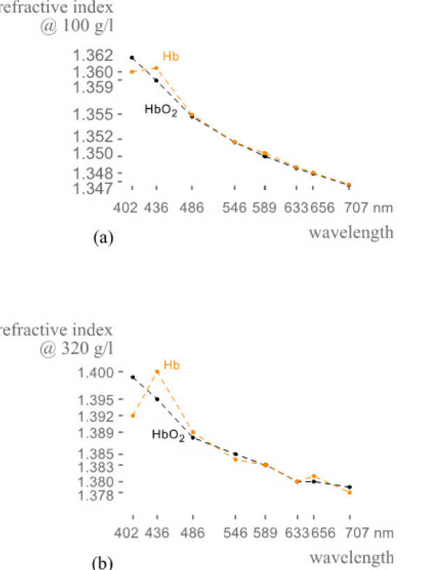
Because the refractive index of hemoglobin in the visible range is sensitive to the hemoglobin concentration, optical investigations of hemoglobin are important for medical diagnostics and treatment. Direct measurements of the refractive index are, however, challenging; few such measurements have previously been reported, especially in a wide wavelength range. We directly measured the refractive index of human deoxygenated and oxygenated hemoglobin for nine wavelengths between 400 and 700 nm for the hemoglobin concentrations up to 140 gl−1. We analyzed the results and suggested a set of model functions to calculate the refractive index depending on the concentration. At all wavelengths, the measured values of the refractive index depended on the concentration linearly. Analyzing the slope of the lines, we determined the specific refraction increments, derived a set of model functions for the refractive index depending on the concentration, and compared our results with those available in the literature. Based on the model functions, we further calculated the refractive index at the physiological concentration within the erythrocytes of 320 gl−1. The results can be used to calculate the refractive index in the visible range for arbitrary concentrations provided that the refractive indices depend on the concentration linearly.
 |
|---|
| Refractive index of oxygenated and deoxygenated haemoglobin (a) measured and (b) calculated at different haemoglobin concentrations |
O. Sydoruk (in collaboration with Erlangen-Nuremberg University and Saratov University)
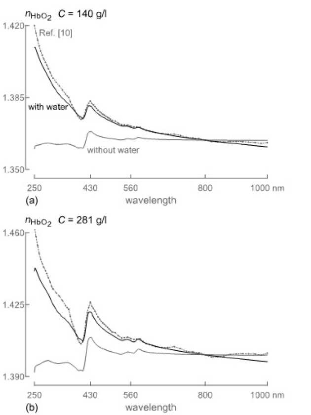
Because direct measurements of the refractive index of hemoglobin over a large wavelength range are challenging, indirect methods deserve particular attention. Among them, the Kramers-Kronig relations are a powerful tool often used to derive the real part of a refractive index from its imaginary part. However, previous attempts to apply the relations to solutions of human hemoglobin have been somewhat controversial, resulting in disagreement between several studies. We showed that this controversy could be resolved when careful attention is paid not only to the absorption of hemoglobin but also to the dispersion of the refractive index of the nonabsorbing solvent. We developed a Kramers-Kroning analysis taking both contributions into account and compare the results with the data from several studies. Good agreement with experiments is found across the visible and parts of near-infrared and ultraviolet regions. These results reinstated the use of the Kramers-Kronig relations for hemoglobin solutions and provide an additional source of information about their refractive index.
 |
|---|
| Comparison between refractive indices calculated using different models at different concentrations. |
Lab-on-a-chip devices
- Rapid Evaporation-driven Chemical Pre-Concentration and Separation on Paper
- MEMS dielectrophoresis device for osteoblast cell stimulation
Richard Syms
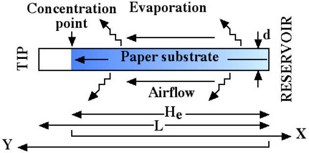
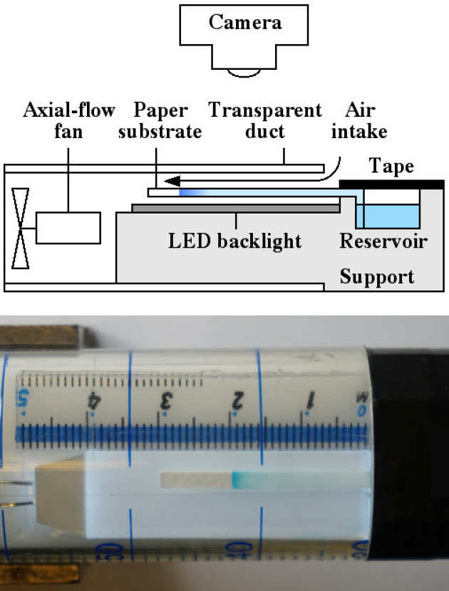
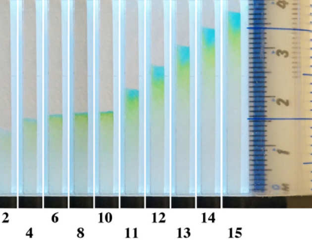
We have investigated airflow-enhanced evaporation as a method for rapid chemical pre-concentration on a thin porous substrate. The mechanism is described by combining 1D models of capillary rise, chromatography and pervaporation concentration. We have shown that the effective length of the column can be shorter than its actual length, allowing concentrate to be held at a stagnation point and then released for separation, and that the Péclet number, which determines concentration performance, is determined only by the substrate properties. We have solved the differential equations dynamically, and shown that faster concentration can be achieved during capillary filling. We have carried out experiments using chromatography paper in a ducted airflow and quantified concentration by optical imaging of water-soluble food dyes. We have obtained good agreement with the model, and achieved concentration factors of 100 in 10 mins using Brilliant Blue FCF. We have demonstrated partial separation of Brilliant Blue from Tartrazine immediately following concentration, on a single un-patterned substrate. The mechanism may provide a method for improving the sensitivity of lab-on-paper devices.
 |
|---|
| Principle of airflow enhanced paper concentration. |
 |
|---|
| Schematic and realisation of apparatus for rapid concentration, showing Brilliant Blue concentrated at an intermediate point along a strip of chromatography paper. |
 |
|---|
| Spatial distribution of Brilliant Blue FCF and Tartrazine at different times T (in minutes) during concentration-separation experiment. |
Helin Zou, Richard Syms,
Steve Mellon and Liz Tanner (Queen Mary University of London)
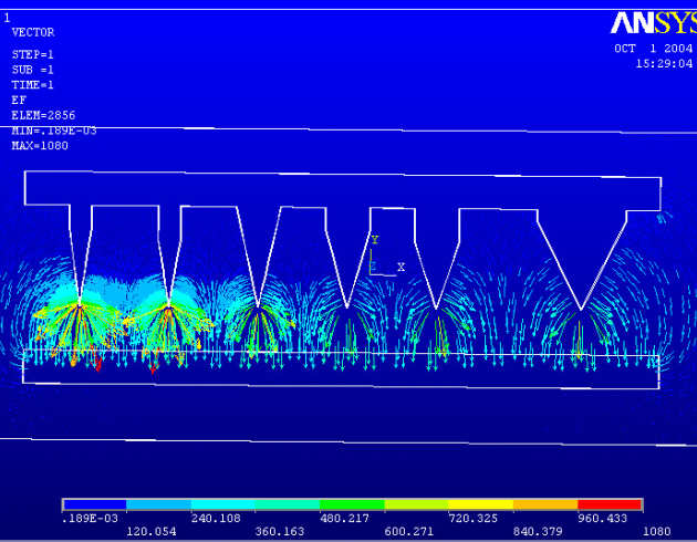
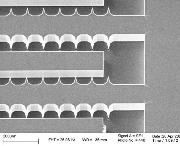
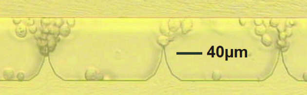
We have developed a fixed microelectrode device for cell stimulation using micro-electro-mechanical systems (MEMS) technology. Dielectrophoretic forces obtained from non-uniform electric fields are used for manipulating and positioning osteoblasts. The experiments show that the osteoblasts experience positive dielectrophoresis (p-DEP) when suspended in iso-osmotic culture medium and exposed to AC fields at 5 MHz frequency. Negative dielectrophoresis (n-DEP) is obtained at 0.1 MHz. The viability of osteoblasts under dielectrophoresis has been investigated. The viability values for cells exposed to DEP are nearly three times higher than the control values, indicating that dielectrophoresis may have an anabolic effect on osteoblasts.
 |
|---|
| ANSYS simulation of dielectrophoretic forces near shaped electrodes |
|
|
|---|
| Prototype dielectrophoretic stimulation device fabricated in BSOI |
 |
|---|
| Osteoblasts concentrated near shaped electrodes by DEP force |