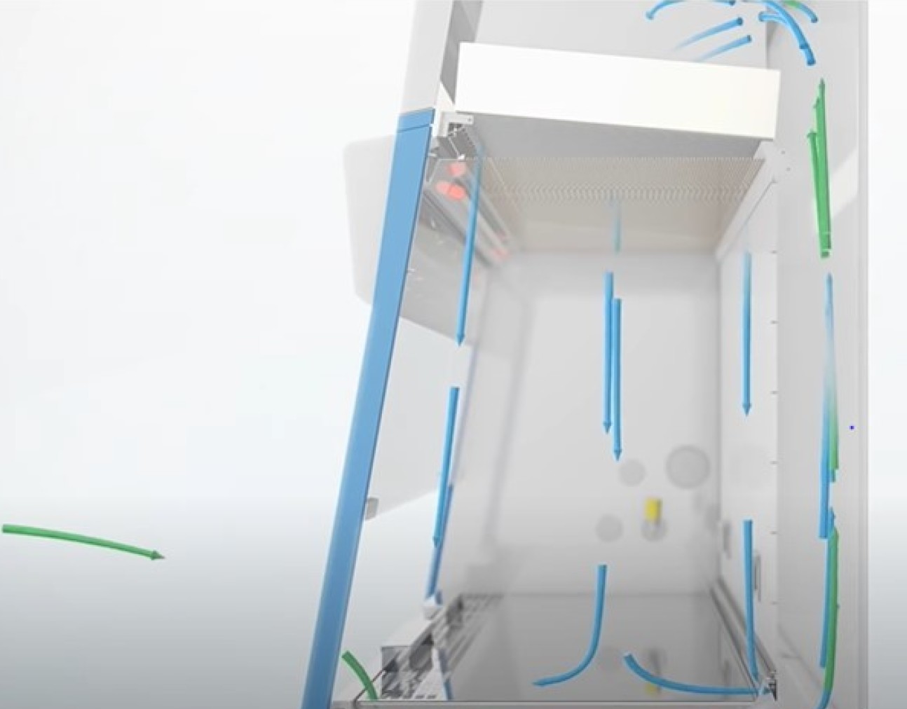
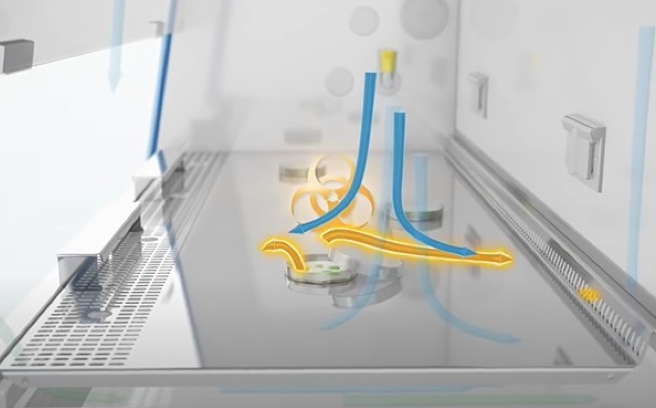
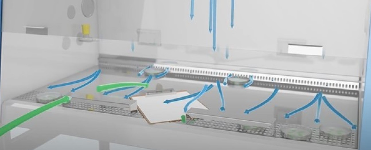
TCimageClass II Facility

An aseptic shared core research facility for all varieties of Cell and Tissue cultures Class I and II, cryopreservation and Bioprinting.

The Facility is designed to accommodate the sterile culture of mammalian cell lines and other animal cell lines, such as insect cells.

Work involving the culture of bacterial cells, spore-forming yeast, Neurospora or fungi or single-celled microorganisms may not be performed due to the potential of these cultures contaminating other cell lines.

All work involving biological material must first be approved by the department and Faculty (class 1 and lower class 2) and also by the safety department for higher Class 2 and above. All work involving recombinant DNA must first be approved by the University GM Committee. All work is reviewed for appropriateness of the work for the Facility and that all protocols / SOPs are completed and potential risks mitigated

TC Facility Essentials

Access: Induction and Training

Access to the facility is granted by the core facility manager upon proof of training, Bio1 risk assessment, knowledge of potential risks, potentially Hepatitis B immunisation, and proficiency in standard and containment level 2 special practices before working with biological agents. On-site induction training in biosafety and standard operational procedures will be provided by the core facility manager.​

  • All users must have an official induction and be signed off before starting work in the facilities.
  • Work cannot commence without an approved Bio1 risk assessment form and relevant SOPs in place.
  • Only users with the ability to demonstrate their understanding and practical aseptic working skills will be given access by the Facility Manager. If any researcher does not work to an appropriate standard to maintain the sterility and classification of the facility, re-training may be required. Continued inability to work to the desired level despite training will result in loss of access. Once your project is finished, you must tell the Facility Manager.
  • It is not allowed to bring unauthorised persons into the cell culture lab.

This process is detailed below:


Training Essentials
Training

Biological Safety - Foundation Training 1. Pdf manual – cell culture basics  1.  Sterile Technique       

 The Fundamentals of Cell Culture​Provided by The Health Protection Agency (note: cost involved)

2. Using a haemocytometer (cell counting)  2.  Passaging cells 
Compressed Gases and Connecting Gas RegulatorsRisk Assessment Foundation Training (RAFT) 3. Understand and Managing Cell Culture Contamination 3.  Freezing cells         
    4.  Thawing cells    ​ 
    5. Cell culture contamination  
        
     
Enter table footer here
Essential for lab training

Lab Rules

  • Get a clean blue lab coat for yourself and keep it in a box with your name. Write your name in a piece of autoclave tape and stick on the lab coat. Do not share lab coats. Do not work in the hood with a guest lab coat.
  • Keep the room tidy, clean and waste-free. Adhere to the rota and communal duties. Do not bring cardboard into the Facility as it is a source of contamination.
  • Never drink, eat, chew gum, use your mobile phone or use earphones in the facilities.
  • Wash your hands with soap and water before and after working in the lab.
  • Research groups must ensure that an experienced researcher is available to train new starters on group specific techniques - to ensure quality, technique and competence. The facility technical manager will always be available to assist and advice.
  • Any contamination must be logged in the available sheets by the door, stating the incubator and hood used and the date. This helps us keep track of potential problems before it becomes widespread.
  •  Any work done in the safety cabinets needs to be booked in advance. If you forget or need to use a free one urgently, book retrospectively as soon as you are done.
  • Flasks and dishes containing contaminated cells are not to be opened within the facility. Seal the container with parafilm and put in the waste bins.
  • Start the airflow in the cabinet at least 2 minutes before working or cleaning.
  • Clean the safety cabinets with distel first and then ethanol before and after using them. Do not just spray, wipe it. Always from back to front.
  • When possible, use the bead bath instead of the water bath.
  •  Make your own aliquots, label them with your name and date. Keep your own bags of flasks, tubes, dishes, etc. Close them with tape and write your name and date.
  • You must regularly check for mycoplasma contamination (maximum 2 month period). Throw away contaminated cultures. You can only work in the mycoplasma-free hoods if your samples tested negative in the last month. If the samples are borderline/positive or unknown, work in a quarantine hood until they are tested negative. Do not bring external equipment into the mycoplasma-free hood, as it cannot be completely sterilised.
  • Avoid talking, singing or whistling when handling TC consumables and especially while using the incubators. Every time you do this, you are spraying mycoplasma and other contaminants on your cells or consumables.
  • Do not double-dip your stripette into any bottles. Use stripettes as much as possible, as opposed to pipettes.
  • Avoid sudden movements while working in the hood. Every time you go out and back in, you introduce contaminated air.
  • Do not open the package of consumables outside of the hood. Also avoid putting packaging and stripettes inside of the hood when possible. Even after spraying with ethanol, introducing non-sterile plastics is a risk of contamination. Open the packages at the entrance of the hood, and with clean gloves take out and put inside the hood only what you need. Keep your stripettes outside if possible and open them individually inside. Avoid any packaging to be in contact with the cabinet surfaces.
  •  Whenever you need to remove a cap or cover, try to avoid putting it down on the work surface. When working with viral or unscreened materials, place the cap facing up. Always recap your bottles, tubes and flasks as soon as possible. The time they remain open should be kept to the minimum necessary.
  •  Spray any equipment or potentially contaminated materials with distel and ethanol and wipe before introducing into the cabinet.
  • Spraying ethanol is not a substitute for good aseptic technique. Most contaminations are due to poor technique and not cross-contamination from others. Try to improve your technique. Do not spray flasks or plates with ethanol.
  • Report to the Facility Manager if you see any person breaking these rules. It is anonymous and it is for everyone’s benefit.
  • All incidents and accidents must be logged on SALUS. 

Health, Safety and Environment

All of our core TC facilities are run as Biosafety Class II, therefore if even your work is lower class you must be aware that others working around you may be working with cells and biological materials that have the potential to cause harm to health.

  • Both B114 and B631a are Class II biological labs. Always be aware that other users might be working with infectious materials. Blood and human tissue should always be handled as if infectious. Viral work (AAV and lentiviral) can only be done in B114. Unscreened human tissue and cells can only be used in B631a.
  • Blue lab coats must be worn in the Cell Culture rooms at all times. White coats are not allowed. Remove your lab coat when exiting the facility. Never use a blue coat in another area. You must ensure that lab coats are regularly laundered by dropping them in the laundry collection point. When a person leaves the Department, the lab coats must be returned for laundering. Please keep a different lab coat for each lab. Do not use anyone else’s lab coat.
  • Gloves must be worn at all times in the cell culture lab, particularly when accessing hoods or incubators. Remember to change them often. Gloves must be removed prior to going out of the cell culture room: when in corridors, do not wear gloves. Transport material inside of close boxes.

Biosafety

Risk Assessment

All work MUST be risk assessed and approved prior to commencing. Each Biological material must have a valid Bio1 risk assessment completed and relevant RADAR record before it may be brought into the facility.

  1. Complete a Bio1 risk assessment form
  2. Send to the facility manager: Miguel Hermida for review
  3. Once agreed this can be uploaded to the RADAR system and an approval process will initiate
  4. Notification of approval will be sent via email, and only once recieved can the biological material be introduced to the facility (following relevant quarantine procedures) and work commence

Equipment

  • Co2 incubators
  • Microscopes
  • Biosafety cabinets

equip

Overview

The Cell / Tissue Culture Facility, is a multi-user laboratory managed and overseen by the departmental dedicated technician \ facility manager (with support from the wider technical team), in the Department of Bioengineering. It is divided in two separate laboratories:

  • Human Primary cell culture: B631a, Bessemer level 6
  • Animal / established/immortalized cell culture / cryopreservation and Bioprinting: B114, level 1 Bessemer.
  • Uren U617 and human Tissue on Uren L3

All researchers using this space must adhere to strict rules on aseptic techniques, including housekeeping standards of care and quality. This is both to protect the users, but is essential for protecting the work - cell cultures are incredibly vulnerable from becoming contaminated from the researchers themselves (including yeasts, bacterias and mycoplasmas).

Aseptic Techniques

Successful cell culture depends heavily on keeping the cells free from contamination by microorganisms such as bacterial, fungi, and viruses.  Nonsterile supplies, media, and reagents, airborne particles laden with microorganisms, unclean incubators, and dirty work surfaces are all sources of biological contamination.

Aseptic technique, designed to provide a barrier between the microrganisms in the environment and the sterile cell culture, depends upon a set of procedures to reduce the probability of contamination from these sources. The elements of aseptic technique are a sterile work area, good personal hygiene, sterile reagents and media, and sterile handling.

View an Aseptic Techniques Checklist

Video: Sterile technique

The steps to prevent contamination of your cell culture and demonstration of best-practice sterile techniques.


Sterile work area

The simplest and most economical way to reduce contamination from airborne particles and aerosols (e.g., dust, spores, shed skin, sneezing) is to use a cell culture hood.  

  • The cell culture hood should be properly set up and be located in an area that is restricted to cell culture that is free from drafts from doors, windows, and other equipment, and with no through traffic.  
  • The work surface should be uncluttered and contain only items required for a particular procedure; it should not be used as a storage area.
  • Before and after use, the work surface should be disinfected thoroughly, and the surrounding areas and equipment should be cleaned routinely.
  • For routine cleaning, wipe the work surface with 70% ethanol before and during work, especially after any spillage.
  • You may use ultraviolet light to sterilize the air and exposed work surfaces in the cell culture hood between uses.
  • Using a Bunsen burner for flaming is not necessary nor is it recommended in a cell culture hood.
  • Leave the cell culture hood running at all times, turning it off only when they will not be used for extended periods of time.

Good personal hygiene

Wash your hands before and after working with cell cultures. 

In addition to protecting you from hazardous materials, wearing personal protective equipment also reduces the probability of contamination from shed skin as well as dirt and dust from your clothes.

Sterile reagents & media

Commercial reagents and media undergo strict quality control to ensure their sterility, but they can become contaminated while handling.  Follow the guidelines below for sterile handling to avoid contaminating them.  Always sterilize any reagents, media, or solutions prepared in the laboratory using the appropriate sterilization procedure (e.g., autoclave, sterile filter).

 
 Sterile handling
  • Always wipe your hands and your work area with 70% ethanol.
  • Wipe the outside of the containers, flasks, plates, and dishes with 70% ethanol before placing them in the cell culture hood. 
  • Avoid pouring media and reagents directly from bottles or flasks.
  • Use sterile glass or disposable plastic pipettes and a pipettor to work with liquids, and use each pipette only once to avoid cross contamination.  Do not unwrap sterile pipettes until they are to be used.  Keep your pipettes at your work area. 
  • Always cap the bottles and flasks after use and seal multi-well plates with tape or place them in resealable bags to prevent microorganisms and airborn contaminants from gaining entry.
  • Never uncover a sterile flask, bottle, petri dish, etc. until the instant you are ready to use it and never leave it open to the environment.  Return the cover as soon as you are finished. 
  • If you remove a cap or cover, and have to put it down on the work surface, place the cap with opening facing down.
  • Use only sterile glassware and other equipment. 
  • Be careful not to talk, sing, or whistle when you are performing sterile procedures. 
  • Perform your experiments as rapidly as possible to minimize contamination.

Biological Safety Cabinet

Biological safety cabinets work by pushing sterile air from the top to the bottom. This is a great tool to maintain sterility if used correctly.

Most contaminations in cell cultures occur when the cabinets are used incorrectly.

 Pic1MSC

Contaminated air (shown in green) is passed through a HEPA filter to deliver a laminar flow of clean air (blue) which is sterile and particle free.

 MSC2


The airflow carries contaminated particles (yellow) down the vents and to the HEPA filters.

 MSC3

Anything placed inside the cabinet will alter the airflow. That is why the cabinet must be as empty as possible and the vents free at all times

3D Bioplotter - Bioprinting

The 3D-Bioplotter System is a versatile rapid prototyping tool for processing a great variety of biomaterials for computer-aided tissue engineering (CATE), from 3D CAD models and patient CT data to the physical 3D scaffold with a designed and defined outer form and an open inner structure.

  • Built-in camera to enhance needle calibration and to ensure consistent prints
  • Temperature controlled build platform and sensor ports. This allows greater material variety and finely tuned environments for low tolerance scaffolding.
  • Five cartridges slots for more materials in a single print.

Further info: EnvisionTEC - YouTube and 3D-Bioplotter® Manufacturer Series | EnvisionTEC

Bioplotter

Cell Culture Contamination

Understanding the Causes and Managing the Risks

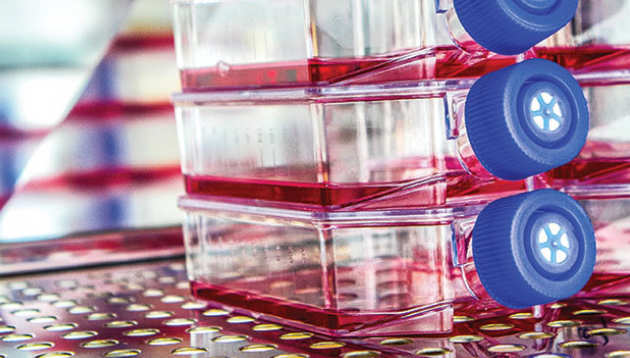
Biological contamination is the dread of every person working with cell culture. When cultures become infected with microorganisms, or cross-contaminated by foreign cells, these cultures usually must be destroyed. Since the sources of culture contamination are ubiquitous as well as difficult to identify and eliminate, no cell culture laboratory remains unaffected by this concern. With the continuing increase in the use of cell culture for biological research, vaccine production, and production of therapeutic proteins for personalized medicine and emerging regenerative medicine applications, culture contamination remains a highly important issue.

Introduction

Cell culture is continuing a 60-year trend of increasing use and importance in academic research, therapeutic medicine, and drug discovery, accompanied by an amplified economic impact.1,2 New therapies, vaccines, and drugs, as well as regenerated and synthetic organs, will increasingly come from cultured mammalian cells. With greater usage and proficiency of cell culture techniques comes a better understanding of the perils and problems associated with cell culture contamination. In the 21st century, there are better testing methods and preventive tools, and an awareness of the risk and effects of contamination requires that cell culturists remain vigilant; undetected contamination can have widespread downstream effects.

Biological contamination: a common companion

 Carolyn Kay Lincoln and Michael Gabridge summed up the contamination problem in 1998 as: “Cell culture contamination continues to be a major problem at the basic research bench as well as for bioproduct manufacturers. Contamination is what truly endangers the use of cell cultures as reliable reagents and tools.”

The biological contamination of mammalian cell cultures is more common than you might think. Statistics reported in the mid-1990s show that between 11 percent and 15 percent of cultures in U.S laboratories were infected with Mycoplasma species. Even with better recognition of the problem and more stringent testing of commercially prepared reagents and media, the incidence of mycoplasma growth in research laboratory cultures was 23 percent in one recent study, and in 2010 an astonishing 8.45 percent of cultures commercially tested from biopharmaceutical sources were contaminated with fungi and bacteria, including mycoplasma.

In the research laboratory, contamination is not just an occasional irritation, but it can cost valuable resources including time and money. Ultimately, contamination can affect the credibility of a research group or particular scientist; publications sometimes must be withdrawn due to fears about retrospective sample contamination or reported results that turn out to be artifacts. In biopharmaceutical manufacturing, contamination can have an even more dramatic effect when entire production runs must be discarded. It is extremely important, therefore, to understand how sample contamination can occur and what methods are available to limit and, ultimately, prevent it.

What causes biological contamination?

Poor Aseptic technique is the number 1 cause of cell culture contamination

Biological contaminants can be divided into two subgroups depending on the ease of detecting them in cultures, with the easiest being most bacteria and fungi. Those that are more difficult to detect, and thus present potentially more serious problems, include Mycoplasmas, viruses, and cross-contamination by other mammalian cells.

Bacteria and fungi

Bacteria and fungi, including molds and yeasts, are ubiquitous in the environment and are able to quickly colonize and flourish in the rich cell culture milieu. Their small size and fast growth rates make these microbes the most commonly encountered cell culture contaminants. In the absence of antibiotics, bacteria can usually be detected in a culture within a few days of contamination, either by microscopic observation or by their direct effects on the culture (pH shifts, turbidity, and cell death). Yeasts generally cause the growth medium to become very cloudy or turbid, whereas molds will produce branched mycelium, which eventually appear as furry clumps floating in the medium.

Mycoplasmas

Mycoplasmas are certainly the most serious and widespread of all the biological contaminants, due to their low detection rates and their effect on mammalian cells. Although mycoplasmas are technically bacteria, they possess certain characteristics that make them unique. They are much smaller than most bacteria (0.15 to 0.3 μm), so they can grow to very high densities without any visible signs. They also lack a cell wall, and that, combined with their small size, means that they can sometimes slip through the pores of filter membranes used in sterilization. Since the most common antibiotics target bacterial cell walls, mycoplasmas are resistant.

Mycoplasmas are extremely detrimental to any cell culture: they affect the host cells’ metabolism and morphology, cause chromosomal aberrations and damage, and can provoke cytopathic responses, rendering any data from contaminated cultures unreliable. In Europe, mycoplasma contamination levels have been found to be extremely high—between 25 percent and 40 percent—and reported rates in Japan have been as high as 80 percent.4 The discrepancy between the U.S. and the rest of the world is likely due to the use of testing programs. Statistics show that laboratories that routinely test for mycoplasma contamination have much lower incidence; once detected, contamination can be contained and eliminated. Testing for mycoplasma should be performed at least once per month, and there is a wide range of commercially available kits. The only way to ensure detection of species is to use at least two different testing methods, such as DAPI staining and PCR.5
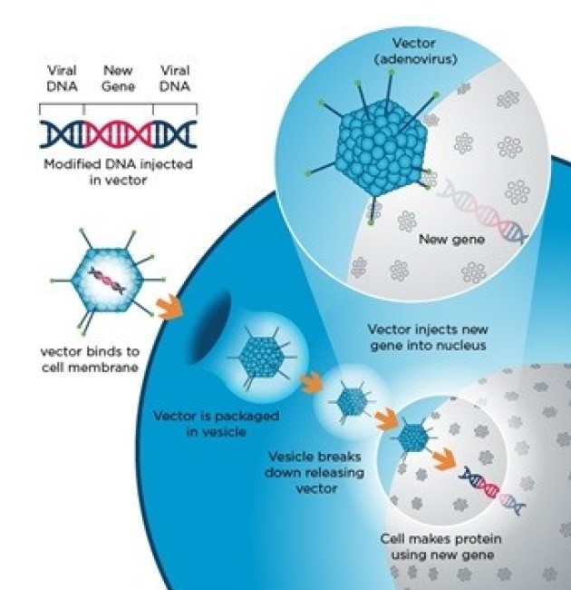
Viruses

Like mycoplasmas, viruses do not provide visual cues to their presence; they do not change the pH of the culture medium or result in turbidity. Since viruses use their host for replication, drugs used to block viruses can also be highly toxic for the cells being cultured. Viruses that cause damage to the host cell do, however, tend to be self-limiting, so the major concern for viral contamination is their potential for infecting laboratory personnel. Those working with human or other primate cells must use extra safety precautions.

Other mammalian cell types

Cross-contamination of a cell culture with other cell types is a serious problem that has only recently been considered alarming.7,8 An estimated 15 percent to 20 percent of cell lines currently in use are misidentified9,10, a problem that began with the first human cell line, HeLa, an unusually aggressive cervical adenocarcinoma isolated from Henrietta Lacks in 1952. HeLa cells are so aggressive that, once accidentally introduced into a culture, they quickly overgrow the original cells. But the problem is not limited to HeLa; there are many examples of cell lines that are characterized as endothelial cells or prostate cancer cells but are actually bladder cancer cells, and characterized as breast cancer cells but are in fact ovarian cancer cells. In these cases, the problem occurs when the foreign cell type is better adapted to the culture conditions, and thus replaces the original cells in the culture. Such contamination clearly poses a problem for the quality of research produced, and the use of cultures containing the wrong cell types can lead to retraction of published results.

Sources of biological contaminants in the lab

In order to reduce the frequency of biological contamination, it is important to understand how biological contaminants can enter culture dishes. In most laboratories, the greatest sources of microbes are those that accompany laboratory personnel. These are circulated as airborne particles and aerosols during normal lab work. Talking, sneezing, and coughing can generate significant amounts of aerosols. Clothing can also harbor and transport a range of microorganisms from outside the lab, so it is crucial to wear lab coats when working in the cell culture lab. Even simply moving around the lab can create air movement, so the room must be cleaned often to reduce dust particles.

Certain laboratory equipment, such as pipetting devices, vortexers, or centrifuges without biocontainment vessels, can generate large amounts of microbial-laden particulates and aerosols. Frequently used laboratory equipment, including water baths, refrigerators, microscopes, and cold storage rooms, are also reservoirs for microbes and fungi. Improperly cleaned and maintained incubators can serve as an acceptable home for fungi and bacteria. Overcrowding of materials in the autoclave during sterilization can also result in incomplete elimination of microbes.

Culture media, bovine sera, reagents, and plasticware can also be major sources of biological contaminants. While commercial testing methods are much improved over those of earlier decades, it is paramount to use materials that are certified for cell culture use. Cross-contamination can occur when working with multiple cell lines at the same time. Each cell type should have its own solutions and supplies and should be manipulated separately from other cells. Unintentional use of nonsterile supplies, media, or solutions during routine cell culture procedures is the major source of microbial spread.

Conclusion

Contamination is a prevalent issue in the culturing of cells, and it is essential that any risks are managed effectively so that experiment integrity is maintained. Antibiotics can be used for a few weeks to ensure resolution of a known microbial contamination; however, routine use should be avoided. Regular inclusion of antibiotics not only selects for resistant organisms, but also masks any low-level infection and habitual mistakes in aseptic technique.

The best approach to fighting contamination is for each person to keep records of all cell culture work including each passage, general cell appearance, and manipulations including feeding, splitting, and counting of cells. If contamination does occur, make a note of the characteristics and the time and date. In this way, any contamination can be pinpointed at the time it occurs and improvements can be made to aseptic techniques or lab protocols. In the next article of this series, we explore in more detail effective measures for contamination prevention, in particular the key role of the CO2 incubator.

Cryostorage

The Department has a -140C freezer available for long term storage of cells and tissues. This is a safer alternative to liquid nitrogen as it removes the need for LN2 exposure. Please contact the Bioscience Technical Team if you need space in the freezer.

Protocol for freezing down cells:

  • Assess cells. They should be healthy and within the logarithmic phase of growth.
  • Harvest cells
  • Prepare the freezing media according to the cell line recommendation
  • Pellet down the desired number of cells as usual
  • Re-suspend in 1ml of freezing media
  • Place in a labelled cryovial. Make sure to add name, cell type and date
  • Place the vials into a Mr Frosty and place it in the -80C freezer for 24h. Make sure the Mr Frosty is at room temperature and the isopropanol level reaches the mark.
  • Transfer to your allocated box in the -140C freezer in Bessemer B125 or UREN615

Virus work

Lentivirs and Adeno associated virus can be used within the core facility, once approved by Bio1 and space has been allocated in the appropriate Viral Incubator and hood

Vectors for Lentiviral Production

Lenti

In addition to normal cell culture facility regulations, workers using viruses (adenovirus or lentivirus) need to follow these points:

  • Only approved and trained persons can work in the virus facilities.
  • DOUBLE gloves and specific cell culture lab coats (green) should be worn at all times. These green lab coats must not be worn outside the cell culture facility. Use safety goggles if there is a risk of splashes to the eyes. 
  • Avoid any procedure that results in production of aerosols, such as aspirating. [If aspirating is absolutely essential the aspirator should be fitted with a filter that blocks viruses such as a hydrophobic HEPA filter]. The aspirator must be decontaminated with Klorcept solution so that the final concentration of Klorcept in the waste is at least 1% Virkon and soaked for minimum 30 min.
  • Avoid use of sharps (needles, glass, metal etc.) whenever possible. If unavoidable, take particular care with handling and disposal: used needles must not be re-sheathed or removed; needles and syringes should always be disposed of as a complete unit into a sharps container (yellow bin).
  • Infectious materials must be transported in sealed primary container inside a sealed and leak-proof secondary containment labelled with a biohazard sticker.
  • All areas in which virus work is done should be sprayed with a 1-5% Klorcept solution (note: solutions must be coloured to be active), followed by 70% ethanol. Note: adenovirus is NOT destroyed by ethanol only, therefore Klorcept is required.
  • Solid waste: all plastic-ware placed inside the cabinet while working with the virus must be decontaminated with Virkon prior to autoclaving in double autoclave bags. This can be done by spraying all plasticware with 1-5% Klorcept solution or by soaking in a 1-5% Klorcept solution. Especially when working with lentivirus, place an autoclave bag in the Class II cabinet; at the end of the session, the waste bag must be sealed before removal from the cabinet, double-bagged, and autoclaved.
  • Liquid waste: treat with 1-5% Klorcept solution for at least 2 hrs before disposing down the sink.
  • Spillages: the area should be sprayed with 1-5% Klorcept solution and 70% ethanol. For larger spills, cover spillage with Klorcept powder and leave for at least 3 min before placing in double biohazard bag for autoclaving. Then clean whole area with 1% Klorcept and 70% ethanol.
  • When centrifuges are used for biologically hazardous materials, safety caps must be used. Rotors must be disinfected with 1% Klorcept solution after each use.