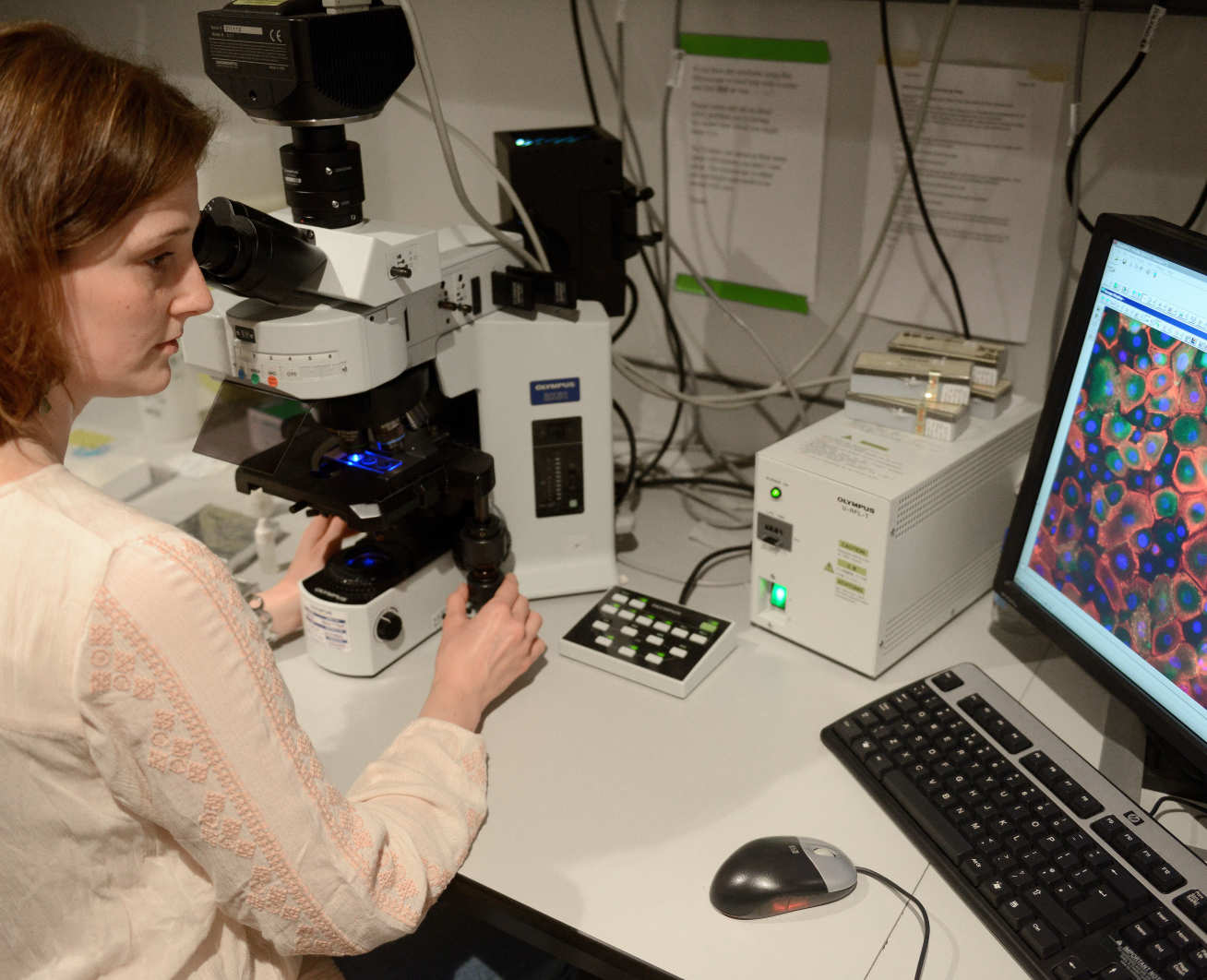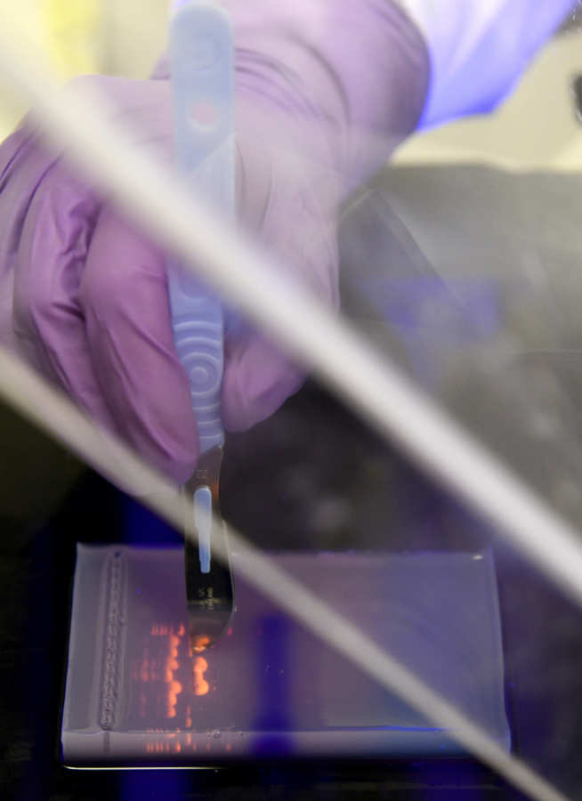

Our research has generated a number of specialised techniques and tools to understand the mechanisms of cell biology processes with impacts on homeostasis and diseases.
Research interests of Dr Vania Braga encompass the use of biosensors to detect localised activation of small GTPases at junctions and other intracellular sites. She has extensive expertise in biochemical and high-throughput methods to determine signalling pathways. The laboratory has also generated a number of image analysis and computer vision tools to quantify distinct phenotypes at junctions caused by manipulation of signalling pathways (Erasmus et al., 2016, 2015; Kelly et al., 2011; Kalaji et al., 2012; Frasa et al., 2010; Lozano et al., 2008).
Dr Anne Burke-Gaffney’s group has set up the xCELLigence Real-Time Cell Analysis (RTCA) system for measuring endothelial barrier function, migration and proliferation. The group also routinely uses the Ibidi Pump System to expose endothelial cells to different flow patterns: continuous unidirectional, oscillating and pulsatile, to simulate blood vessel conditions.
Dr Charlotte Dean’s group has established a new method to visualise the process of alveolar formation live, in slices of lung tissue. This system will enable us to test signalling mechanisms and potential treatments to repair damaged alveoli (Akram et al. 2019, Nature Communications 10:1178).
Dr Michael Emerson’s laboratory investigates human platelet aggregation in vitro using blood samples from volunteers and patients in collaboration with Chelsea and Westminster Hospital. A bench-top flow cytometer allows mechanistic investigation of platelet activation. The lab also has expertise in mouse models in vivo (Tymvios C, Jones S, Moore C, Pitchford SC, Page CP, Emerson M 2008). Real-time measurement of non-lethal platelet thromboembolic responses in the anaesthetized mouse. Thromb. Haemost. 99, 435-40) and in working with nanoparticles.
Dr Nicholas Kirkby’s group uses conditional knockout in vivo models and classical pharmacological bioassays to understand how interactions between the vascular wall and platelets control thrombotic risk. A particular focus is how these processes are influenced by aspirin and non-steroidal anti-inflammatory drugs to provide anti-thrombotic efficacy or reduce cardiovascular toxicity.
Dr Birgit Leitinger’s group are developing FRET-based sensors to understand the molecular mechanism of receptor activation in collaboration with Paul French’s group (Department of Physics, Faculty of Natural Sciences). The group uses fusion proteins with mCherry or GFP, as well as SNAP-tagged receptors which are labelled with a mixture of red- and green-fluorescent cell-impermeable dyes.
Professor Jane Mitchell’s group utilizes a range of vascular bioassay techniques including novel assays combining autologous vascular and immune cells to construct ‘same donor’ pharmacological platforms.
Dr Gregory Quinlan’s group in collaboration with Professor Alan Spivey’s group, Imperial College Chemistry is developing an innovative system to limit post-operative acute kidney injury and related morbidities in patients subject to cardiac surgery involving cardiopulmonary diseases. It involves a bypass inline filter to bind and remove key products of haemolysis (haemoglobin, haem and iron) from extracorporeal circuits.