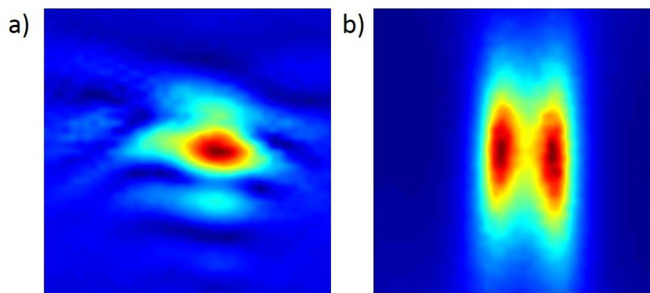
Ultrasound imaging using Full Matrix Capture (FMC) has brought a step change to the capabilities of phased arrays for the detection and characterisation of defects for Non-Destructive Evaluation (NDE). The majority of the algorithms that are used to process the FMC measured data are based on the so-called Beam-Forming (BF) approach. This has a theoretical limit of resolution that has long been held to be the best possible for imaging. However, recent research in a variety of application areas (e.g. medical) has shown that this theoretical resolution limit may in fact be surpassed by alternative processing of the measured data. This is based on the extraction of additional information in the signals related to the interaction of waves between multiple objects scatterers. Such new approaches have become known as Super Resolution (SR) algorithms. The images in Figure 1, constructed from experimental measurements, illustrate this potential very clearly.
The research project will develop these SR algorithms further and apply them to defect sizing for ultrasonic NDE applied to safety critical components in the nuclear industry. The application focus will be achieved by centring the research on the specific, but commonly arising, problem of the detection and characterisation of a defect at or adjacent to the back wall of a component. The challenge here is that the reflected signal from the back wall can overwhelm the reflections from the defect which are often much smaller, for example the diffracted signal from a crack tip. The resolution can be improved so as to separate these signal components by raising the frequency, but then difficulties are encountered due to scattering noise in the base material. Thus the aim of the project is to develop improved imaging methods to:
- Optimise the detection and characterisation of a small defect at or near the back surface of a component, using an ultrasonic array at the front surface.
- Characterise properties of "difficult" host materials, so enabling improved contrast of defect images.
- Define the basis for the selection of frequency so as to optimise detection and characterisation of such defects in difficult materials.
Funding Sources and Sponsor
EPSRC EP/M016315/1 (Centre for Doctoral Training in Non-Destructive Evaluation), Rolls-Royce plc, Amec Foster Wheeler plc.