Overview
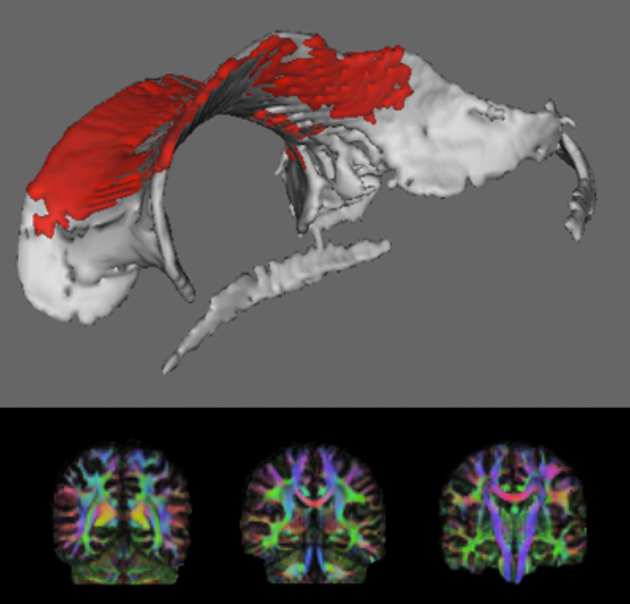
 Traumatic brain injury (TBI) is the most common cause of death and disability in young adults. It is most commonly caused by road traffic accidents and assaults. Patients can experience problems with concentration, attention span, and memory. This cognitive impairment poses an immense burden to well-being, and leads to significant economic and social consequences. In military personnel, TBI may occur following blast injury, which has become the signature injury from the current conflicts in Iraq and Afghanistan. Approximately 25% of TBI patients improve but an equal number deteriorate over time. We know little about why patients vary so much in how they recover. Crucially, we have no treatments to improve cognitive impairment and brain recovery after TBI. When we are injured our bodies mount an inflammatory response to start the healing process. This may be helpful initially but if this process continues it can be damaging. Recently, using a Positron Emission Tomography (PET) scan, abnormal persistent brain inflammation – chronic neuroinflammation – has been detected following TBI. Little is known about how chronic neuroinflammation following TBI relates to brain recovery. We will use two types of brain scan to investigate chronic neuroinflammation and brain recovery: PET and Magnetic Resonance Imaging (MRI). PET scans allow us to measure inflammation directly. MRI scans measure brain structure and function. We will assess psychological and social recovery. We are also investigating whether particular genes lead to poor outcomes and whether the metabolic syndrome, a condition causing long-term inflammation in the body, also is associated with chronic neuroinflammation.
Principal investigator
Researchers involved
- Claire Feeney
- Amy Jolly
Funding
- Medical Research Council